
Page 641 - Textbook of Pathology, 6th Edition
P. 641
625
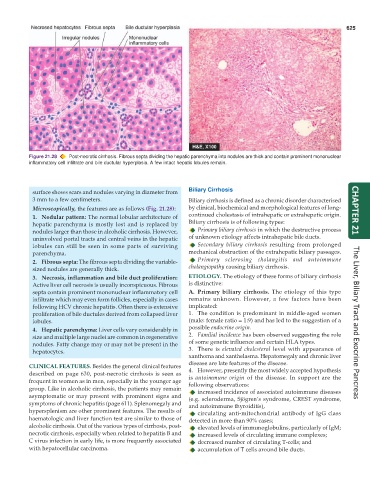
Figure 21.28 Post-necrotic cirrhosis. Fibrous septa dividing the hepatic parenchyma into nodules are thick and contain prominent mononuclear
inflammatory cell infiltrate and bile ductular hyperplasia. A few intact hepatic lobules remain.
surface shows scars and nodules varying in diameter from Biliary Cirrhosis
3 mm to a few centimeters. Biliary cirrhosis is defined as a chronic disorder characterised
Microscopically, the features are as follows (Fig. 21.28): by clinical, biochemical and morphological features of long- CHAPTER 21
1. Nodular pattern: The normal lobular architecture of continued cholestasis of intrahepatic or extrahepatic origin.
hepatic parenchyma is mostly lost and is replaced by Biliary cirrhosis is of following types:
nodules larger than those in alcoholic cirrhosis. However, Primary biliary cirrhosis in which the destructive process
uninvolved portal tracts and central veins in the hepatic of unknown etiology affects intrahepatic bile ducts.
lobules can still be seen in some parts of surviving Secondary biliary cirrhosis resulting from prolonged
parenchyma. mechanical obstruction of the extrahepatic biliary passages.
2. Fibrous septa: The fibrous septa dividing the variable- Primary sclerosing cholangitis and autoimmune
sized nodules are generally thick. cholangiopathy causing biliary cirrhosis.
3. Necrosis, inflammation and bile duct proliferation: ETIOLOGY. The etiology of these forms of biliary cirrhosis
Active liver cell necrosis is usually inconspicuous. Fibrous is distinctive:
septa contain prominent mononuclear inflammatory cell A. Primary biliary cirrhosis. The etiology of this type
infiltrate which may even form follicles, especially in cases remains unknown. However, a few factors have been
following HCV chronic hepatitis. Often there is extensive implicated:
proliferation of bile ductules derived from collapsed liver 1. The condition is predominant in middle-aged women
lobules. (male: female ratio = 1:9) and has led to the suggestion of a The Liver, Biliary Tract and Exocrine Pancreas
4. Hepatic parenchyma: Liver cells vary considerably in possible endocrine origin.
size and multiple large nuclei are common in regenerative 2. Familial incidence has been observed suggesting the role
nodules. Fatty change may or may not be present in the of some genetic influence and certain HLA types.
hepatocytes. 3. There is elevated cholesterol level with appearance of
xanthoma and xanthelasma. Hepatomegaly and chronic liver
disease are late features of the disease.
CLINICAL FEATURES. Besides the general clinical features
described on page 630, post-necrotic cirrhosis is seen as 4. However, presently the most widely accepted hypothesis
is autoimmune origin of the disease. In support are the
frequent in women as in men, especially in the younger age following observations:
group. Like in alcoholic cirrhosis, the patients may remain increased incidence of associated autoimmune diseases
asymptomatic or may present with prominent signs and (e.g. scleroderma, Sjögren’s syndrome, CREST syndrome,
symptoms of chronic hepatitis (page 611). Splenomegaly and and autoimmune thyroiditis),
hypersplenism are other prominent features. The results of circulating anti-mitochondrial antibody of IgG class
haematologic and liver function test are similar to those of detected in more than 90% cases;
alcoholic cirrhosis. Out of the various types of cirrhosis, post- elevated levels of immunoglobulins, particularly of IgM;
necrotic cirrhosis, especially when related to hepatitis B and increased levels of circulating immune complexes;
C virus infection in early life, is more frequently associated decreased number of circulating T-cells; and
with hepatocellular carcinoma. accumulation of T cells around bile ducts.