
Page 638 - Textbook of Pathology, 6th Edition
P. 638
622
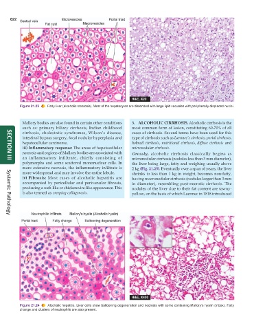
Figure 21.23 Fatty liver (alcoholic steatosis). Most of the hepatocytes are distended with large lipid vacuoles with peripherally displaced nuclei.
Mallory bodies are also found in certain other conditions 3. ALCOHOLIC CIRRHOSIS. Alcoholic cirrhosis is the
such as: primary biliary cirrhosis, Indian childhood most common form of lesion, constituting 60-70% of all
cirrhosis, cholestatic syndromes, Wilson’s disease, cases of cirrhosis. Several terms have been used for this
intestinal bypass surgery, focal nodular hyperplasia and type of cirrhosis such as Laennec’s cirrhosis, portal cirrhosis,
hepatocellular carcinoma. hobnail cirrhosis, nutritional cirrhosis, diffuse cirrhosis and
iii) Inflammatory response: The areas of hepatocellular micronodular cirrhosis.
necrosis and regions of Mallory bodies are associated with Grossly, alcoholic cirrhosis classically begins as
an inflammatory infiltrate, chiefly consisting of micronodular cirrhosis (nodules less than 3 mm diameter),
SECTION III
polymorphs and some scattered mononuclear cells. In the liver being large, fatty and weighing usually above
more extensive necrosis, the inflammatory infiltrate is 2 kg (Fig. 21.25). Eventually over a span of years, the liver
more widespread and may involve the entire lobule. shrinks to less than 1 kg in weight, becomes non-fatty,
iv) Fibrosis: Most cases of alcoholic hepatitis are having macronodular cirrhosis (nodules larger than 3 mm
accompanied by pericellular and perivenular fibrosis, in diameter), resembling post-necrotic cirrhosis. The
producing a web-like or chickenwire-like appearance. This nodules of the liver due to their fat content are tawny-
is also termed as creeping collagenosis. yellow, on the basis of which Laennec in 1818 introduced
Systemic Pathology
Figure 21.24 Alcoholic hepatitis. Liver cells show ballooning degeneration and necrosis with some containing Mallory’s hyalin (Inbox). Fatty
change and clusters of neutrophils are also present.