
Page 639 - Textbook of Pathology, 6th Edition
P. 639
623
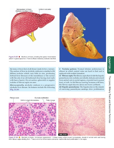
Figure 21.25 Alcoholic cirrhosis, showing the typical micronodular
pattern in gross specimen. There is diffuse nodularity (nodules less than
3 mm diameter) on sectioned surface of the liver.
the term cirrhosis first of all (from Greek kirrhos = tawny). i) Nodular pattern: Normal lobular architecture is CHAPTER 21
The surface of liver in alcoholic cirrhosis is studded with effaced in which central veins are hard to find and is
diffuse nodules which vary little in size, producing replaced with nodule formation.
hobnail liver (because of the resemblance of the surface ii) Fibrous septa: The fibrous septa that divide the hepatic
with the sole of an old-fashioned shoe having short nails parenchyma into nodules are initially delicate and extend
with heavy heads). On cut section, spheroidal or angular from central vein to portal regions, or portal tract to portal
nodules of fibrous septa are seen. tract, or both. As the fibrous scarring increases with time,
Microscopically, alcoholic cirrhosis is a progressive the fibrous septa become dense and more confluent.
alcoholic liver disease. Its features include the following iii) Hepatic parenchyma: The hepatocytes in the islands
(Fig. 21.26): of surviving parenchyma undergo slow proliferation The Liver, Biliary Tract and Exocrine Pancreas
Figure 21.26 Alcoholic cirrhosis, microscopic appearance. It shows nearly uniform-sized micronodules, devoid of central veins and having
thick fibrous septa dividing them. There is minimal inflammation and some reactive bile duct proliferation in the septa.