
Page 643 - Textbook of Pathology, 6th Edition
P. 643
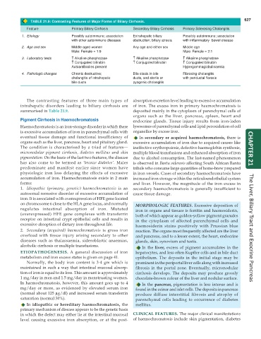
TABLE 21.9: Contrasting Features of Major Forms of Biliary Cirrhosis. 627
Feature Primary Biliary Cirrhosis Secondary Biliary Cirrhosis Primary Sclerosing Cholangitis
1. Etiology Possibly autoimmune; association Extrahepatic biliary Possibly autoimmune; association
with other autoimmune diseases obstruction; biliary atresia with inflammatory bowel disease
2. Age and sex Middle-aged women Any age and either sex Middle age
Male: Female = 1:9 Male: Female = 2:1
3. Laboratory tests ↑ ↑ ↑ ↑ ↑ Alkaline phosphatase ↑ ↑ ↑ ↑ ↑ Alkaline phosphatase ↑ ↑ ↑ ↑ ↑ Alkaline phosphatase
↑ ↑ ↑ ↑ ↑ Conjugated bilirubin ↑ ↑ ↑ ↑ ↑ Conjugated bilirubin ↑ ↑ ↑ ↑ ↑ Conjugated bilirubin
Autoantibodies present Hypergammaglobulinaemia
4. Pathologic changes Chronic destructive; Bile stasis in bile Fibrosing cholangitis
cholangitis of intrahepatic ducts, and sterile or with periductal fibrosis
bile ducts pyogenic cholangitis
The contrasting features of three main types of absorption excretion level leading to excessive accumulation
intrahepatic disorders leading to biliary cirrhosis are of iron. The excess iron in primary haemochromatosis is
summarised in Table 21.9. deposited mainly in the cytoplasm of parenchymal cells of
organs such as the liver, pancreas, spleen, heart and
Pigment Cirrhosis in Haemochromatosis endocrine glands. Tissue injury results from iron-laden
Haemochromatosis is an iron-storage disorder in which there lysosomes of parenchymal cells and lipid peroxidation of cell
is excessive accumulation of iron in parenchymal cells with organelles by excess iron.
eventual tissue damage and functional insufficiency of In secondary or acquired haemochromatosis, there is
organs such as the liver, pancreas, heart and pituitary gland. excessive accumulation of iron due to acquired causes like
The condition is characterised by a triad of features— ineffective erythropoiesis, defective haemoglobin synthesis, CHAPTER 21
micronodular pigment cirrhosis, diabetes mellitus and skin multiple blood transfusions and enhanced absorption of iron
pigmentation. On the basis of the last two features, the disease due to alcohol consumption. The last-named phenomenon
has also come to be termed as ‘bronze diabetes’. Males is observed in Bantu siderosis affecting South African Bantu
predominate and manifest earlier since women have tribals who consume large quantities of home-brew prepared
physiologic iron loss delaying the effects of excessive in iron vessels. Cases of secondary haemochromatosis have
accumulation of iron. Haemochromatosis exists in 2 main increased iron storage within the reticuloendothelial system
forms: and liver. However, the magnitude of the iron excess in
1. Idiopathic (primary, genetic) haemochromatosis is an secondary haemochromatosis is generally insufficient to
autosomal recessive disorder of excessive accumulation of cause tissue damage.
iron. It is associated with overexpression of HFE gene located
on chromosome 6 close to the HLA gene locus, and normally MORPHOLOGIC FEATURES. Excessive deposition of
regulates intestinal absorption of iron. Mutated iron in organs and tissues is ferritin and haemosiderin,
(overexpressed) HFE gene complexes with transferrin both of which appear as golden-yellow pigment granules
receptor on intestinal crypt epithelial cells and results in in the cytoplasm of affected parenchymal cells and
excessive absoption of dietary iron throughout life. haemosiderin stains positively with Prussian blue
2. Secondary (acquired) haemochromatosis is gross iron reaction. The organs most frequently affected are the liver The Liver, Biliary Tract and Exocrine Pancreas
overload with tissue injury arising secondary to other and pancreas, and to a lesser extent, the heart, endocrine
diseases such as thalassaemia, sideroblastic anaemias, glands, skin, synovium and testis.
alcoholic cirrhosis or multiple transfusions. In the liver, excess of pigment accumulates in the
ETIOPATHOGENESIS. A general discussion of iron hepatocytes, and less often Kupffer cells and in bile duct
metabolism and iron excess states is given on page 41. epithelium. The deposits in the initial stage may be
Normally, the body iron content is 3-4 gm which is prominent in the periportal liver cells along with increased
maintained in such a way that intestinal mucosal absorp- fibrosis in the portal zone. Eventually, micronodular
tion of iron is equal to its loss. This amount is approximately cirrhosis develops. The deposits may produce grossly
1 mg/day in men and 1.5 mg/day in menstruating women. chocolate-brown colour of the liver and nodular surface.
In haemochromatosis, however, this amount goes up to 4 In the pancreas, pigmentation is less intense and is
mg/day or more, as evidenced by elevated serum iron found in the acinar and islet cells. The deposits in pancreas
(normal about 125 μg/dl) and increased serum transferrin produce diffuse interstitial fibrosis and atrophy of
saturation (normal 30%). parenchymal cells leading to occurrence of diabetes
In idiopathic or hereditary haemochromatosis, the mellitus.
primary mechanism of disease appears to be the genetic basis
in which the defect may either lie at the intestinal mucosal CLINICAL FEATURES. The major clinical manifestations
level causing excessive iron absorption, or at the post- of haemochromatosis include skin pigmentation, diabetes