
Page 642 - Textbook of Pathology, 6th Edition
P. 642
626 B. Secondary biliary cirrhosis. Most cases of secondary
biliary cirrhosis result from prolonged obstruction of
extrahepatic biliary passages (page 599). These causes
include the following:
1. Extrahepatic cholelithiasis, most common
2. Biliary atresia
3. Cancer of biliary tree and of head of pancreas
4. Postoperative strictures with superimposed ascending
cholangitis.
C. Cirrhosis due to primary sclerosing cholangitis. Primary
or idiopathic sclerosing cholangitis is a chronic cholestatic
syndrome of unknown etiology. It is characterised by
progressive, inflammatory, sclerosing and obliterative
process affecting the entire biliary passages, both extra-
hepatic and intrahepatic ducts. Although etiology remains
unknown, various mechanisms have been postulated which
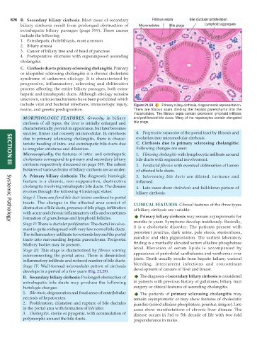
include viral and bacterial infections, immunologic injury, Figure 21.29 Primary biliary cirrhosis, diagrammatic representation.
toxins, and genetic predisposition. There are fibrous scars dividing the hepatic parenchyma into the
micronodules. The fibrous septa contain prominent lymphoid infiltrate
MORPHOLOGIC FEATURES. Grossly, in biliary and proliferated bile ducts. Many of the hepatocytes contain elongated
cirrhosis of all types, the liver is initially enlarged and bile plugs.
characteristically greenish in appearance, but later becomes
smaller, firmer and coarsely micronodular. In cirrohosis 4. Progressive expansion of the portal tract by fibrosis and
due to primary sclerosing cholangitis, there is charac- evolution into micronodular cirrhosis.
teristic beading of intra- and extrahepatic bile ducts due C. Cirrhosis due to primary sclerosing cholangitis:
to irregular strictures and dilatation. Following changes are seen:
Microscopically, the features of intra- and extrahepatic 1. Fibrosing cholangitis with lymphocytic infiltrate around
cholestasis correspond to primary and secondary biliary bile ducts with segmental involvement.
cirrhosis respectively discussed on page 599. The salient 2. Periductal fibrosis with eventual obliteration of lumen
SECTION III
features of various forms of biliary cirrhosis are as under: of affected bile ducts.
A. Primary biliary cirrhosis: The diagnostic histologic 3. Intervening bile ducts are dilated, tortuous and
feature is a chronic, non-suppurative, destructive inflamed.
cholangitis involving intrahepatic bile ducts. The disease 4. Late cases show cholestasis and full-blown picture of
evolves through the following 4 histologic states: biliary cirrhosis.
Stage I: There are florid bile duct lesions confined to portal
tracts. The changes in the affected area consist of CLINICAL FEATURES. Clinical features of the three types
destruction of bile ducts, presence of bile plugs, infiltration of biliary cirrhosis are variable:
with acute and chronic inflammatory cells and sometimes
formation of granulomas and lymphoid follicles. Primary biliary cirrhosis may remain asymptomatic for
Systemic Pathology
Stage II: There is ductular proliferation. The ductal involve- months to years. Symptoms develop insidiously. Basically,
ment is quite widespread with very few normal bile ducts. it is a cholestatic disorder. The patients present with
The inflammatory infiltrate too extends beyond the portal persistent pruritus, dark urine, pale stools, steatorrhoea,
tracts into surrounding hepatic parenchyma. Periportal jaundice and skin pigmentation. The earliest laboratory
Mallory bodies may be present. finding is a markedly elevated serum alkaline phosphatase
level. Elevation of serum lipids is accompanied by
Stage III: This stage is characterised by fibrous scarring appearance of periorbital xanthelasma and xanthomas over
interconnecting the portal areas. There is diminished
inflammatory infiltrate and reduced number of bile ducts. joints. Death usually results from hepatic failure, variceal
Stage IV: Well-formed micronodular pattern of cirrhosis bleeding, intercurrent infections and concomitant
develops in a period of a few years (Fig. 21.29). development of cancers of liver and breast.
B. Secondary biliary cirrhosis: Prolonged obstruction of The diagnosis of secondary biliary cirrhosis is considered
extrahepatic bile ducts may produce the following in patients with previous history of gallstones, biliary tract
histologic changes: surgery or clinical features of ascending cholangitis.
1. Bile stasis, degeneration and focal areas of centrilobular The patients of primary sclerosing cholangitis may
necrosis of hepatocytes. remain asymptomatic or may show features of cholestatic
2. Proliferation, dilatation and rupture of bile ductules jaundice (raised alkaline phosphatase, pruritus, fatigue). Late
in the portal area with formation of bile lakes. cases show manifestations of chronic liver disease. The
3. Cholangitis, sterile or pyogenic, with accumulation of disease occurs in 3rd to 5th decade of life with two fold
polymorphs around the bile ducts. preponderance in males.