
Page 657 - Textbook of Pathology, 6th Edition
P. 657
641
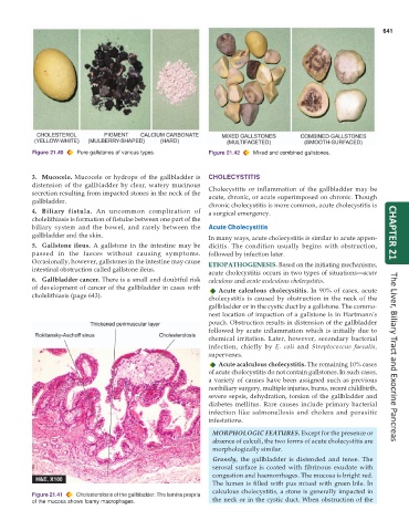
Figure 21.40 Pure gallstones of various types. Figure 21.42 Mixed and combined gallstones.
3. Mucocele. Mucocele or hydrops of the gallbladder is CHOLECYSTITIS
distension of the gallbladder by clear, watery mucinous Cholecystitis or inflammation of the gallbladder may be
secretion resulting from impacted stones in the neck of the acute, chronic, or acute superimposed on chronic. Though
gallbladder.
chronic cholecystitis is more common, acute cholecystitis is
4. Biliary fistula. An uncommon complication of a surgical emergency.
cholelithiasis is formation of fistulae between one part of the
biliary system and the bowel, and rarely between the Acute Cholecystitis
gallbladder and the skin. In many ways, acute cholecystitis is similar to acute appen- CHAPTER 21
5. Gallstone ileus. A gallstone in the intestine may be dicitis. The condition usually begins with obstruction,
passed in the faeces without causing symptoms. followed by infection later.
Occasionally, however, gallstones in the intestine may cause ETIOPATHOGENESIS. Based on the initiating mechanisms,
intestinal obstruction called gallstone ileus.
acute cholecystitis occurs in two types of situations—acute
6. Gallbladder cancer. There is a small and doubtful risk calculous and acute acalculous cholecystitis.
of development of cancer of the gallbladder in cases with Acute calculous cholecystitis. In 90% of cases, acute
cholelithiasis (page 643). cholecystitis is caused by obstruction in the neck of the
gallbladder or in the cystic duct by a gallstone. The commo-
nest location of impaction of a gallstone is in Hartmann’s
pouch. Obstruction results in distension of the gallbladder
followed by acute inflammation which is initially due to
chemical irritation. Later, however, secondary bacterial
infection, chiefly by E. coli and Streptococcus faecalis,
supervenes. The Liver, Biliary Tract and Exocrine Pancreas
Acute acalculous cholecystitis. The remaining 10% cases
of acute cholecystitis do not contain gallstones. In such cases,
a variety of causes have been assigned such as previous
nonbiliary surgery, multiple injuries, burns, recent childbirth,
severe sepsis, dehydration, torsion of the gallbladder and
diabetes mellitus. Rare causes include primary bacterial
infection like salmonellosis and cholera and parasitic
infestations.
MORPHOLOGIC FEATURES. Except for the presence or
absence of calculi, the two forms of acute cholecystitis are
morphologically similar.
Grossly, the gallbladder is distended and tense. The
serosal surface is coated with fibrinous exudate with
congestion and haemorrhages. The mucosa is bright red.
The lumen is filled with pus mixed with green bile. In
calculous cholecystitis, a stone is generally impacted in
Figure 21.41 Cholesterolosis of the gallbladder. The lamina propria
of the mucosa shows foamy macrophages. the neck or in the cystic duct. When obstruction of the