
Page 652 - Textbook of Pathology, 6th Edition
P. 652
636
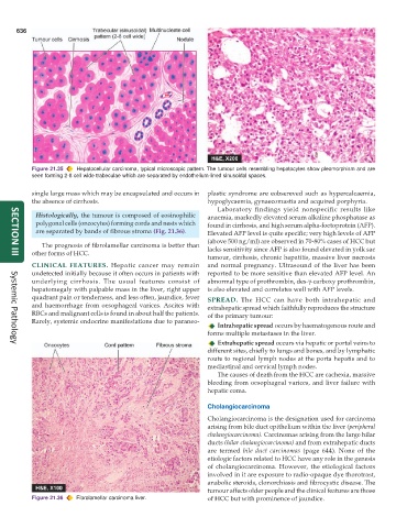
Figure 21.35 Hepatocellular carcinoma, typical microscopic pattern. The tumour cells resembling hepatocytes show pleomorphism and are
seen forming 2-8 cell wide trabeculae which are separated by endothelium-lined sinusoidal spaces.
single large mass which may be encapsulated and occurs in plastic syndrome are eobsereved such as hypercalcaemia,
the absence of cirrhosis. hypoglycaemia, gynaecomastia and acquired porphyria.
Laboratory findings yield nonspecific results like
Histologically, the tumour is composed of eosinophilic anaemia, markedly elevated serum alkaline phosphatase as
polygonal cells (oncocytes) forming cords and nests which found in cirrhosis, and high serum alpha-foetoprotein (AFP).
are separated by bands of fibrous stroma (Fig. 21.36). Elevated AFP level is quite specific; very high levels of AFP
(above 500 ng/ml) are observed in 70-80% cases of HCC but
The prognosis of fibrolamellar carcinoma is better than
other forms of HCC. lacks sensitivity since AFP is also found elevated in yolk sac
tumour, cirrhosis, chronic hepatitis, massive liver necrosis
SECTION III
CLINICAL FEATURES. Hepatic cancer may remain and normal pregnancy. Ultrasound of the liver has been
undetected initially because it often occurs in patients with reported to be more sensitive than elevated AFP level. An
underlying cirrhosis. The usual features consist of abnormal type of prothrombin, des-γ-carboxy prothrombin,
hepatomegaly with palpable mass in the liver, right upper is also elevated and correlates well with AFP levels.
quadrant pain or tenderness, and less often, jaundice, fever SPREAD. The HCC can have both intrahepatic and
and haemorrhage from oesophageal varices. Ascites with extrahepatic spread which faithfully reproduces the structure
RBCs and malignant cells is found in about half the patients. of the primary tumour:
Rarely, systemic endocrine manifestations due to paraneo-
Intrahepatic spread occurs by haematogenous route and
forms multiple metastases in the liver.
Extrahepatic spread occurs via hepatic or portal veins to
Systemic Pathology
different sites, chiefly to lungs and bones, and by lymphatic
route to regional lymph nodes at the porta hepatis and to
mediastinal and cervical lymph nodes.
The causes of death from the HCC are cachexia, massive
bleeding from oesophageal varices, and liver failure with
hepatic coma.
Cholangiocarcinoma
Cholangiocarcinoma is the designation used for carcinoma
arising from bile duct epithelium within the liver (peripheral
cholangiocarcinoma). Carcinomas arising from the large hilar
ducts (hilar cholangiocarcinoma) and from extrahepatic ducts
are termed bile duct carcinomas (page 644). None of the
etiologic factors related to HCC have any role in the genesis
of cholangiocarcinoma. However, the etiological factors
involved in it are exposure to radio-opaque dye thorotrast,
anabolic steroids, clonorchiasis and fibrocystic disease. The
tumour affects older people and the clinical features are those
Figure 21.36 Fibrolamellar carcinoma liver. of HCC but with prominence of jaundice.