
Page 653 - Textbook of Pathology, 6th Edition
P. 653
637
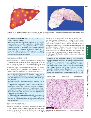
Figure 21.37 Metastatic tumour deposits in the liver as seen on sectioned surface. Characteristic features include multiple, variable-sized,
nodular masses, often under the capsule, producing umbilication on the surface.
MORPHOLOGIC FEATURES. Grossly, the tumour is frequent primary tumours metastasising to the liver, in
firm to hard and whitish. descending order of frequency, are those of stomach, breast,
Microscopically, the tumour has glandular structure. The lungs, colon, oesophagus, pancreas, malignant melanoma
tumour cells resemble biliary epithelium but without bile and haematopoietic malignancies. Sarcomas rarely
secretion. They form various patterns such as tubular, metastasise to the liver. Occasionally, metastatic involvement
ductular or papillary. The stroma consists of fibrous tissue may be present in the absence of obvious evidence of primary
with little or no capillary formation. Occasionally, tumour. Aside from general features of disseminated
mucinous, signet-ring and adenosquamous type of malignancy such as anorexia, cachexia and anaemia, the
patterns are found. An uncommon variant is combined patients have hepatomegaly with nodular free margin. There CHAPTER 21
hepatocellular-cholangiocarcinoma. is little hepatic dysfunction until late in the course of hepatic
metastatic disease.
Hepatoblastoma (Embryoma) MORPHOLOGIC FEATURES. Grossly, most metastatic
Hepatoblastoma is a rare malignant tumour arising from carcinomas form multiple, spherical, nodular masses
primitive hepatic parenchymal cells. It presents before the which are of variable size. Liver is enlarged and heavy,
age of 2 years as progressive abdominal distension with weighing 5 kg or more. The tumour deposits are white,
anorexia, failure to thrive, fever and jaundice. It is more well-demarcated, soft or haemorrhagic. The surface of the
common in boys. The concentration of serum AFP is high. liver shows characteristic umbilication due to central
The tumour grows rapidly and causes death by necrosis of nodular masses (Fig. 21.37).
haemorrhage, hepatic failure or widespread metastases. Histologically, the metastatic tumours generally
reproduce the structure of the primary lesions (Fig. 21.38).
MORPHOLOGIC FEATURES, Grossly, the tumour is
circumscribed and lobulated mass measuring 5-25 cm in
size, having areas of cystic degeneration, haemorrhage
and necrosis. The Liver, Biliary Tract and Exocrine Pancreas
Microscopically, hepatoblastoma consists of 2
components:
i) Epithelial component contains 2 types of cells—
‘embryonal’ hepatocytes are small with dark-staining,
hyperchromatic nuclei and scanty cytoplasm, and ‘foetal’
hepatocytes are larger with more cytoplasm that may be
granular or clear. The epithelial cells are organised in
trabeculae, ribbons or rosettes.
ii) Mesenchymal component includes fibrous connective
tissue, cartilage and osteoid of variable degree of
maturation. Extramedullary haematopoiesis is a frequent
accompaniment.
Secondary Hepatic Tumours
Metastatic tumours in the liver are more common than the
primary hepatic tumours. Most frequently, they are blood-
borne metastases, irrespective of whether the primary Figure 21.38 Metastatic deposits from undifferentiated sarcoma in
tumour is drained by portal vein or systemic veins. Most the liver.