
Page 658 - Textbook of Pathology, 6th Edition
P. 658
642
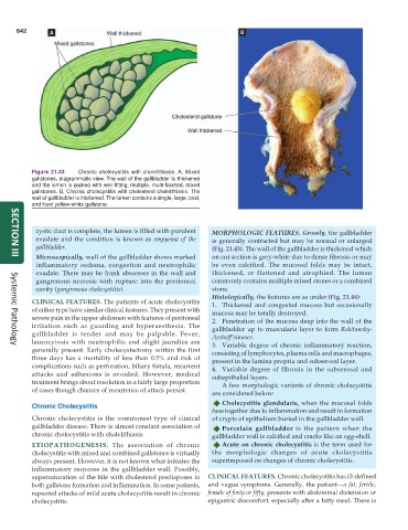
Figure 21.43 Chronic cholecystitis with cholelithiasis. A, Mixed
gallstones, diagrammatic view. The wall of the gallbladder is thickened
and the lumen is packed with well-fitting, multiple, multi-faceted, mixed
gallstones. B, Chronic cholecystitis with cholesterol cholelithiasis. The
wall of gallbladder is thickened. The lumen contains a single, large, oval,
and hard yellow-white gallstone.
cystic duct is complete, the lumen is filled with purulent MORPHOLOGIC FEATURES. Grossly, the gallbladder
exudate and the condition is known as empyema of the is generally contracted but may be normal or enlarged
gallbladder. (Fig. 21.43). The wall of the gallbladder is thickened which
Microscopically, wall of the gallbladder shows marked on cut section is grey-white due to dense fibrosis or may
SECTION III
inflammatory oedema, congestion and neutrophilic be even calcified. The mucosal folds may be intact,
exudate. There may be frank abscesses in the wall and thickened, or flattened and atrophied. The lumen
gangrenous necrosis with rupture into the peritoneal commonly contains multiple mixed stones or a combined
cavity (gangrenous cholecystitis). stone.
Histologically, the features are as under (Fig. 21.44):
CLINICAL FEATURES. The patients of acute cholecystitis 1. Thickened and congested mucosa but occasionally
of either type have similar clinical features. They present with mucosa may be totally destroyed.
severe pain in the upper abdomen with features of peritoneal 2. Penetration of the mucosa deep into the wall of the
irritation such as guarding and hyperaesthesia. The
gallbladder is tender and may be palpable. Fever, gallbladder up to muscularis layer to form Rokitansky-
leucocytosis with neutrophilia and slight jaundice are Aschoff’sinuses.
3. Variable degree of chronic inflammatory reaction,
Systemic Pathology
generally present. Early cholecystectomy within the first consisting of lymphocytes, plasma cells and macrophages,
three days has a mortality of less than 0.5% and risk of present in the lamina propria and subserosal layer.
complications such as perforation, biliary fistula, recurrent 4. Variable degree of fibrosis in the subserosal and
attacks and adhesions is avoided. However, medical subepithelial layers.
treatment brings about resolution in a fairly large proportion A few morphologic variants of chronic cholecystitis
of cases though chances of recurrence of attack persist.
are considered below:
Cholecystitis glandularis, when the mucosal folds
Chronic Cholecystitis
fuse together due to inflammation and result in formation
Chronic cholecystitis is the commonest type of clinical of crypts of epithelium buried in the gallbladder wall.
gallbladder disease. There is almost constant association of Porcelain gallbladder is the pattern when the
chronic cholecystitis with cholelithiasis. gallbladder wall is calcified and cracks like an egg-shell.
ETIOPATHOGENESIS. The association of chronic Acute on chronic cholecystitis is the term used for
cholecystitis with mixed and combined gallstones is virtually the morphologic changes of acute cholecystitis
always present. However, it is not known what initiates the superimposed on changes of chronic cholecystitis.
inflammatory response in the gallbladder wall. Possibly,
supersaturation of the bile with cholesterol predisposes to CLINICAL FEATURES. Chronic cholecystitis has ill-defined
both gallstone formation and inflammation. In some patients, and vague symptoms. Generally, the patient—a fat, fertile,
repeated attacks of mild acute cholecystitis result in chronic female of forty or fifty, presents with abdominal distension or
cholecystitis. epigastric discomfort, especially after a fatty meal. There is