
Page 673 - Textbook of Pathology, 6th Edition
P. 673
neoplastic cystic lesions of the kidney are described later 657
(page 694).
I. Multicystic Renal Dysplasia
The term ‘multicystic renal dysplasia’ or Potter type II is used
for disorganised metanephrogenic differentiation with
persistence of structures in the kidney which are not
represented in normal nephrogenesis. Renal dysplasia is the
most common form of cystic renal disease in the newborn
and infants. The condition may occur sporadically or maybe
familial and part of a syndrome of other anomalies. It is
commonly associated with obstructive abnormalities of the
ureter and lower urinary tract such as obstruction of
pelviureteric junction (PUJ), ureteral atresia and urethral
obstruction.
MORPHOLOGIC FEATURES. Renal dysplasia may be
unilateral or bilateral. The dysplastic process may involve
the entire renal mass or a part of it.
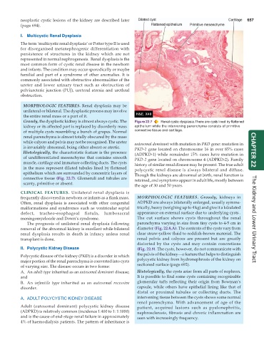
Grossly, the dysplastic kidney is almost always cystic. The Figure 22.7 Renal cystic dysplasia. There are cysts lined by flattened
kidney or its affected part is replaced by disorderly mass epithelium while the intervening parenchyma consists of primitive
of multiple cysts resembling a bunch of grapes. Normal connective tissue and cartilage.
renal parenchyma is almost totally obscured by the mass
while calyces and pelvis may not be recognised. The ureter autosomal dominant with mutation in PKD gene: mutation in
is invariably abnormal, being either absent or atretic. PKD-1 gene located on chromosome 16 in over 85% cases
Histologically, the characteristic feature is the presence (ADPKD-1) while remainder 15% cases have mutation in CHAPTER 22
of undifferentiated mesenchyme that contains smooth PKD-2 gene located on chromosome 4 (ADPKD-2). Family
muscle, cartilage and immature collecting ducts. The cysts history of similar renal disease may be present. The true adult
in the mass represent dilated tubules lined by flattened polycystic renal disease is always bilateral and diffuse.
epithelium which are surrounded by concentric layers of Though the kidneys are abnormal at birth, renal function is
connective tissue (Fig. 22.7). Glomeruli and tubules are retained, and symptoms appear in adult life, mostly between
scanty, primitive or absent. the age of 30 and 50 years.
CLINICAL FEATURES. Unilateral renal dysplasia is
frequently discovered in newborn or infants as a flank mass. MORPHOLOGIC FEATURES. Grossly, kidneys in
Often, renal dysplasia is associated with other congenital ADPKD are always bilaterally enlarged, usually symme-
malformations and syndromes such as ventricular septal trically, heavy (weighing up to 4 kg) and give it a lobulated
defect, tracheo-esophageal fistula, lumbosacral appearance on external surface due to underlying cysts .
meningomyelocele and Down’s syndrome. The cut surface shows cysts throughout the renal The Kidney and Lower Urinary Tract
The prognosis of unilateral renal dysplasia following parenchyma varying in size from tiny cysts to 4-5 cm in
removal of the abnormal kidney is excellent while bilateral diameter (Fig. 22.8,A). The contents of the cysts vary from
renal dysplasia results in death in infancy unless renal clear straw-yellow fluid to reddish-brown material. The
transplant is done. renal pelvis and calyces are present but are greatly
distorted by the cysts and may contain concretions
II. Polycystic Kidney Disease (Fig. 22.9). The cysts, however, do not communicate with
Polycystic disease of the kidney (PKD) is a disorder in which the pelvis of the kidney—a feature that helps to distinguish
major portion of the renal parenchyma is converted into cysts polycystic kidney from hydronephrosis of the kidney on
of varying size. The disease occurs in two forms: sectioned surface (page 692).
A. An adult type inherited as an autosomal dominant disease; Histologically, the cysts arise from all parts of nephron.
and It is possible to find some cysts containing recognisable
glomerular tufts reflecting their origin from Bowman’s
B. An infantile type inherited as an autosomal recessive
disorder. capsule, while others have epithelial lining like that of
distal or proximal tubules or collecting ducts. The
A. ADULT POLYCYSTIC KIDNEY DISEASE intervening tissue between the cysts shows some normal
renal parenchyma. With advancement of age of the
Adult (autosomal dominant) polycystic kidney disease patient, acquired lesions such as pyelonephritis,
(ADPKD) is relatively common (incidence 1:400 to 1: 1:1000) nephrosclerosis, fibrosis and chronic inflammation are
and is the cause of end-stage renal failure in approximately seen with increasingly frequency.
4% of haemodialysis patients. The pattern of inheritance is