
Page 674 - Textbook of Pathology, 6th Edition
P. 674
658 berry aneurysms of the circle of Willis. Any acquired renal
disease is more prone to occur in polycystic kidneys.
B. INFANTILE POLYCYSTIC KIDNEY DISEASE
The infantile (autosomal recessive) form of polycystic kidney
disease (ARPKD) is distinct from the adult form and is less
common (incidence 1:20,000 births). It is transmitted as an
autosomal recessive trait and the family history of similar
disease is usually not present. The condition occurs due to a
mutation in chromosome 6—6p21, PKHD1 (polycystic kidney
and hepatic disease 1). It is invariably bilateral. The age at
presentation may be perinatal, neonatal, infantile or juvenile,
but frequently serious manifestations are present at birth and
result in death from renal failure in early childhood.
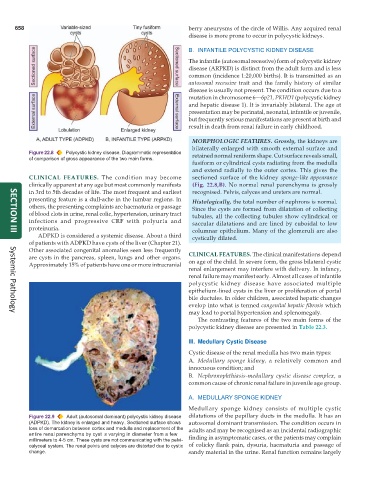
MORPHOLOGIC FEATURES. Grossly, the kidneys are
bilaterally enlarged with smooth external surface and
Figure 22.8 Polycystic kidney disease. Diagrammatic representation retained normal reniform shape. Cut surface reveals small,
of comparison of gross appearance of the two main forms.
fusiform or cylindrical cysts radiating from the medulla
and extend radially to the outer cortex. This gives the
CLINICAL FEATURES. The condition may become sectioned surface of the kidney sponge-like appearance
clinically apparent at any age but most commonly manifests (Fig. 22.8,B). No normal renal parenchyma is grossly
in 3rd to 5th decades of life. The most frequent and earliest recognised. Pelvis, calyces and ureters are normal.
presenting feature is a dull-ache in the lumbar regions. In Histologically, the total number of nephrons is normal.
others, the presenting complaints are haematuria or passage Since the cysts are formed from dilatation of collecting
of blood clots in urine, renal colic, hypertension, urinary tract tubules, all the collecting tubules show cylindrical or
infections and progressive CRF with polyuria and saccular dilatations and are lined by cuboidal to low
proteinuria. columnar epithelium. Many of the glomeruli are also
ADPKD is considered a systemic disease. About a third cystically dilated.
SECTION III
of patients with ADPKD have cysts of the liver (Chapter 21).
Other associated congenital anomalies seen less frequently
are cysts in the pancreas, spleen, lungs and other organs. CLINICAL FEATURES. The clinical manifestations depend
Approximately 15% of patients have one or more intracranial on age of the child. In severe form, the gross bilateral cystic
renal enlargement may interfere with delivery. In infancy,
renal failure may manifest early. Almost all cases of infantile
polycystic kidney disease have associated multiple
epithelium-lined cysts in the liver or proliferation of portal
bile ductules. In older children, associated hepatic changes
evelop into what is termed congenital hepatic fibrosis which
may lead to portal hypertension and splenomegaly.
Systemic Pathology
The contrasting features of the two main forms of the
polycystic kidney disease are presented in Table 22.3.
III. Medullary Cystic Disease
Cystic disease of the renal medulla has two main types:
A. Medullary sponge kidney, a relatively common and
innocuous condition; and
B. Nephronophthiasis-medullary cystic disease complex, a
common cause of chronic renal failure in juvenile age group.
A. MEDULLARY SPONGE KIDNEY
Medullary sponge kidney consists of multiple cystic
Figure 22.9 Adult (autosomal dominant) polycystic kidney disease dilatations of the papillary ducts in the medulla. It has an
(ADPKD). The kidney is enlarged and heavy. Sectioned surface shows autosomal dominant transmission. The condition occurs in
loss of demarcation between cortex and medulla and replacement of the adults and may be recognised as an incidental radiographic
entire renal parenchyma by cyst s varying in diameter from a few finding in asymptomatic cases, or the patients may complain
millimeters to 4-5 cm. These cysts are not communicating with the pelvi-
calyceal system. The renal pelvis and calyces are distorted due to cystic of colicky flank pain, dysuria, haematuria and passage of
change. sandy material in the urine. Renal function remains largely