
Page 676 - Textbook of Pathology, 6th Edition
P. 676
660 GLOMERULAR DISEASES underlying condition. A firm diagnosis, however, can be
established by examination of renal biopsy under light,
DEFINITION AND CLASSIFICATION electron and immunofluorescence microscopy.
Glomerular diseases encompass a large and clinically A number of clinical syndromes are recognised in
significant group of renal diseases. Glomerulonephritis (GN) glomerular diseases. The following are six major glomerular
or Bright’s disease is the term used for diseases that primarily syndromes commonly found in different glomerular diseases:
involve the renal glomeruli. It is convenient to classify nephritic and nephrotic syndromes;
glomerular diseases into 2 broad groups: acute and chronic renal failure;
I. Primary glomerulonephritis in which the glomeruli are the asymptomatic proteinuria and haematuria.
predominant site of involvement. These are briefly described below.
II. Secondary glomerular diseases include certain systemic and I. ACUTE NEPHRITIC SYNDROME. This is the acute
hereditary diseases which secondarily affect the glomeruli. onset of haematuria, proteinuria, hypertension, oedema and
Though this division is widely followed, it is somewhat
arbitrary since many primary forms of glomerulonephritis oliguria following an infective illness about 10 to 20 days
have systemic effects, and many systemic diseases may earlier.
initially present with glomerular involvement. Many 1. The haematuria is generally slight giving the urine smoky
classifications of different types of glomerulonephiritis have appearance and erythrocytes are detectable by microscopy
been described, but most widely accepted classification is or by chemical testing for haemoglobin. Appearance of red
based on clinical presentation and pathologic changes in the cell casts is another classical feature of acute nephritic
glomeruli given in Table 22.4. syndrome.
2. The proteinuria is mild (less than 3 gm per 24 hrs) and is
CLINICAL MANIFESTATIONS usually non-selective (nephritic range proteinuria).
3. Hypertension is variable depending upon the severity
The clinical presentation of glomerular disease is quite
variable but in general four features—proteinuria, haema- of the glomerular disease but is generally mild.
turia, hypertension and disturbed excretory function, are 4. Oedema in nephritic syndrome is usually mild and
present in varying combinations depending upon the results from sodium and water retention (page 97).
5. Oliguria is variable and reflects the severity of glomerular
SECTION III
involvement.
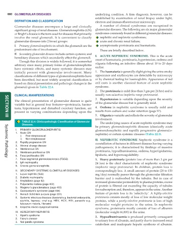
TABLE 22.4: Clinicopathologic Classification of Glomerular The underlying causes of acute nephritic syndrome may
Diseases.
be primary glomerulonephritic diseases (classically acute
I. PRIMARY GLOMERULONEPHRITIS glomerulonephritis and rapidly progressive glomerulo-
1. Acute GN nephritis) or certain systemic diseases (Table 22.5).
i) Post-streptococcal
ii) Non-streptococcal II. NEPHROTIC SYNDROME Nephrotic syndrome is a
2. Rapidly progressive GN constellation of features in different diseases having varying
3. Minimal change disease pathogenesis; it is characterised by findings of massive
4. Membranous GN proteinuria, hypoalbuminaemia, oedema, hyperlipidaemia,
5. Membrano-proliferative GN
6. Focal proliferative GN lipiduria, and hypercoagulability.
7. Focal segmental glomerulosclerosis (FSGS) 1. Heavy proteinuria (protein loss of more than 3 gm per
Systemic Pathology
8. IgA nephropathy 24 hrs) is the chief characteristic of nephrotic syndrome
9. Chronic glomerulonephritis
(nephrotic range proteinuria). In children, protein loss is
II. SECONDARY SYSTEMIC GLOMERULAR DISEASES correspondingly less. A small amount of protein (20 to 150
1. Lupus nephritis (SLE) mg/day) normally passes through the glomerular filtration
2. Diabetic nephropathy barrier and is reabsorbed by the tubules. But in case of
3. Amyloidosis (page 82) increased glomerular permeability to plasma proteins, excess
4. Polyarteritis nodosa (page 402) of protein is filtered out exceeding the capacity of tubules
5. Wegener’s granulomatosis (page 403) for reabsorption and, therefore, appears in the urine. Another
6. Goodpasture’s syndrome (page 494)
7. Henoch-Schönlein purpura (page 331) feature of protein loss is its ‘selectivity’. A highly-selective
8. Systemic infectious diseases (bacterial e.g. bacterial endocarditis, proteinuria consists mostly of loss of low molecular weight
syphilis, leprosy; viral e.g. HBV, HCV, HIV; parasitic e.g. proteins, while a poorly-selective proteinuria is loss of high
falciparum malaria, filariasis) molecular weight proteins in the urine. In nephrotic
9. Idiopathic mixed cryoglobulinaemia
syndrome, proteinuria mostly consists of loss of albumin
III. HEREDITARY NEPHRITIS (molecular weight 66,000) in the urine.
1. Alport’s syndrome 2. Hypoalbuminaemia is produced primarily consequent
2. Fabry’s disease to urinary loss of albumin, and partly due to increased renal
3. Nail-patella syndrome
catabolism and inadequate hepatic synthesis of albumin.