
Page 675 - Textbook of Pathology, 6th Edition
P. 675
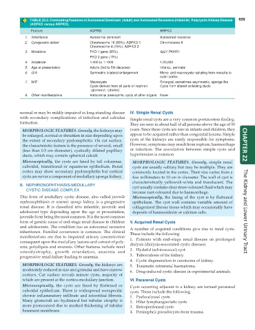
TABLE 22.3. Contrasting Features of Autosomal Dominant (Adult) and Autosomal Recessive (Infantile) Polycystic Kidney Disease 659
(ADPKD versus ARPKD).
Feature ADPKD ARPKD
1. Inheritance Autosomal dominant Autosomal recessive
2. Cytogenetic defect Chromosome 16 (85%): ADPKD-1 Chromosome 6
Chromosome 4 (15%): ADPKD-2
3. Mutations PKD 1 gene (85%) 6p21 PKHD1
PKD 2 gene (15%)
4. Incidence 1:400 to 1:1000 1:20,000
5. Age at presentation Adults (3rd to 5th decades) Infancy, perinatal
6. G/A Symmetric bilateral enlargement Micro- and macrocysts radiating from medulla to
outer cortex
7. M/E Macrocysts Enlarged, sometimes asymmetric, sponge-like
Cysts derived from all parts of nephron Cysts from dilated collecting ducts
(glomeruli, tubules)
8. Other manifestations Intracranial aneurysms, cysts of other organs None
normal or may be mildly impaired in long-standing disease IV. Simple Renal Cysts
with secondary complications of infection and calculus Simple renal cysts are a very common postmortem finding.
formation. They are seen in about half of all persons above the age of 50
MORPHOLOGIC FEATURES. Grossly, the kidneys may years. Since these cysts are rare in infants and children, they
be enlarged, normal or shrunken in size depending upon appear to be acquired rather than congenital lesions. Simple
the extent of secondary pyelonephritis. On cut surface, cysts of the kidneys are rarely responsible for symptoms.
the characteristic feature is the presence of several, small However, symptoms may result from rupture, haemorrhage CHAPTER 22
(less than 0.5 cm diameter), cystically dilated papillary or infection. The association between simple cysts and
ducts, which may contain spherical calculi. hypertension is common.
Microscopically, the cysts are lined by tall columnar, MORPHOLOGIC FEATURES. Grossly, simple renal
cuboidal, transitional or squamous epithelium. Renal cysts are usually solitary but may be multiple. They are
cortex may show secondary pyelonephritis but cortical commonly located in the cortex. Their size varies from a
cysts are never a component of medullary sponge kidney. few millimeters to 10 cm in diameter. The wall of cyst is
characteristically yellowish-white and translucent. The
B. NEPHRONOPHTHIASIS-MEDULLARY cyst usually contains clear straw-coloured fluid which may
CYSTIC DISEASE COMPLEX
become rust-coloured due to haemorrhage.
This form of medullary cystic disease, also called juvenile Microscopically, the lining of the cyst is by flattened
nephronophthiasis or uraemic sponge kidney, is a progressive epithelium. The cyst wall contains variable amount of
renal disease. It is classified into infantile, juvenile and collagenised fibrous tissue which may occasionally have
adolescent type depending upon the age at presentation, deposits of haemosiderin or calcium salts. The Kidney and Lower Urinary Tract
juvenile form being the most common. It is the most common
form of genetic cause of end-stage renal disease in children V. Acquired Renal Cysts
and adolescents. The condition has an autosomal recessive A number of acquired conditions give rise to renal cysts.
inheritance. Familial occurrence is common. The clinical These include the following:
manifestations are due to impaired urinary concentration 1. Patients with end-stage renal disease on prolonged
consequent upon the medullary lesions and consist of poly- dialysis (dialysis-associated cystic disease).
uria, polydipsia and enuresis. Other features include renal
osteodystrophy, growth retardation, anaemia and 2. Hydatid (echinococcal) cyst.
progressive renal failure leading to uraemia. 3. Tuberculosis of the kidney.
4. Cystic degeneration in carcinoma of kidney.
MORPHOLOGIC FEATURES. Grossly, the kidneys are 5. Traumatic intrarenal haematoma.
moderately reduced in size and granular and have narrow 6. Drug-induced cystic disease in experimental animals.
cortices. Cut surface reveals minute cysts, majority of
which are present at the cortico-medullary junction. VI. Pararenal Cysts
Microscopically, the cysts are lined by flattened or Cysts occurring adjacent to a kidney are termed pararenal
cuboidal epithelium. There is widespread nonspecific cysts. These include the following:
chronic inflammatory infiltrate and interstitial fibrosis. 1. Pyelocalyceal cysts
Many glomeruli are hyalinised but tubular atrophy is 2. Hilar lymphangiectatic cysts
more pronounced due to marked thickening of tubular 3. Retroperitoneal cysts
basement membrane. 4. Perinephric pseudocysts from trauma.