
Page 678 - Textbook of Pathology, 6th Edition
P. 678
662
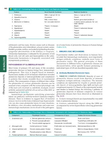
TABLE 22.7: Contrasting Features of Acute Nephritic and Nephrotic Syndromes.
Feature Acute Nephritic Syndrome Nephrotic Syndrome
1. Proteinuria Mild (< 3 gm per 24 hrs) Heavy (> 3 gm per 24 hrs)
2. Hypoalbuminaemia Uncommon Present
3. Oedema Mild, in loose tissue Marked, generalised peripheral
+
+
4. Mechanism of Oedema Na and water retention ↓ ↓ ↓ ↓ ↓ plasma osmotic pressure, Na and
water retention
5. Haematuria Present, microscopic Absent
6. Hypertension Present Present in advanced disease
7. Hyperlipidaemia Absent Present
8. Lipiduria Absent Present
9. Oliguria Present Present in advanced disease
10. Hypercoagulability Absent Present
adolescents and has many diverse causes such as diseases sis of some forms of glomerular diseases in human beings
of the glomerulus, renal interstitium, calyceal system, ureter, (Table 22.9).
bladder, prostate, urethra, and underlying bleeding disorder,
congenital abnormalities of the kidneys or neoplasia. I. IMMUNOLOGIC MECHANISMS
Glomerular haematuria is indicated by the presence of red Experimental studies and observations in humans have
blood cells, red cell casts and haemoglobin in the urine. revealed that immunologic mechanisms, most importantly
Glomerular haematuria is frequently associated with antigen-antibody complexes, underlie most forms of
asymptomatic proteinuria.
glomerular injury. The general principles of these
mechanisms in different forms of glomerular diseases are
PATHOGENESIS OF GLOMERULAR INJURY
discussed below, while specific features pertaining to
Most forms of primary GN and many of the secondary individual types of GN are described separately later.
glomerular diseases in human beings have immunologic
SECTION III
pathogenesis. This view is largely based on immuno- A. Antibody-Mediated Glomerular Injury
fluorescence studies of GN in humans which have revealed
glomerular deposits of immunoglobulins and complement 1. IMMUNE COMPLEX DISEASE. Majority of cases of
in patterns that closely resemble those of experimental glomerular disease result from deposits of immune
models. The consequences of injury at different sites within complexes (antigen-antibody complexes). The immune
the glomerulus in various glomerular diseases can be complexes are represented by irregular or granular glomer-
assessed when compared with the normal physiologic role ular deposits of immunoglobulins (IgG, IgM and IgA) and
of the main cells involved i.e. endothelial, mesangial, visceral complement (mainly C3). Based on the experimental models
epithelial, and parietal epithelial cells as well as of the GBM as and studies in human beings, the following 3 patterns of
summed up in Table 22.8. glomerular deposits of immune complexes in various
Immunologic mechanisms underlying glomerular glomerular diseases have been observed as illustrated in
Systemic Pathology
injury are primarily antibody-mediated (immune-complex Fig. 22.10:
disease). There is evidence to suggest that cell-mediated i) Exclusive mesangial deposits are characterised by very mild
immune reactions in the form of delayed type hypersensiti- form of glomerular disease.
vity can also cause glomerular injury in some situations. ii) Extensive subendothelial deposits along the GBM are
In addition, a few secondary mechanisms and some non- accompanied by severe hypercellular sclerosing glomerular
immunologic mechanisms are involved in the pathogene- lesions.
TABLE 22.8: Relationship of Physiologic Role of Glomerular Components with Consequences in Glomerular Injury.
Component Physiologic Function Consequence of Injury Related Glomerular Disease
1. Endothelial cells i) Maintain glomerular perfusion Vasoconstriction Acute renal failure
ii) Prevent leucocyte adhesion Leucocyte infiltration Focal/diffuse proliferative GN
iii) Prevent platelet aggregation Intravascular microthrombi Thrombotic microangiopathies
2. Mesangial cells Control glomerular filtration Proliferation and increased matrix Membranoproliferative GN
3. Visceral epithelial cells Prevent plasma protein filtration Proteinuria Minimal change disease, FSGS
4. GBM Prevents plasma protein filtration Proteinuria Membranous GN, ? MPGN
5. Parietal epithelial cells Maintain Bowman’s space Crescent formation RPGN