
Page 725 - Textbook of Pathology, 6th Edition
P. 725
TUMOUR MARKERS. Germ cell tumours of the testis 45 years. Testicular germ cell tumours are almost always 709
secrete polypeptide hormones and certain enzymes which malignant. Nearly half of them contain more than one
can be detected in the blood. Two tumour markers widely histologic type. Germ cell tumours are also found at the
used in the diagnosis, staging and monitoring the follow-up extragonadal sites such as the retroperitoneum and media-
of patients with testicular tumours are: human chorionic stinum, besides their counterparts in the female gonads (page
gonadotropin (hCG) and alpha-foetoprotein (AFP). In 745).
addition, carcinoembryonic antigen (CEA), human placental
lactogen (HPL), placental alkaline phosphatase, testosterone, Intratubular Germ Cell Neoplasia
oestrogen and luteinising hormone may also be elevated. The term intratubular germ cell neoplasia (ITGCN) is used
hCG is synthesised by placental syncytio-trophoblast to describe the preinvasive stage of germ cell tumours,
such as in various non-seminomatous germ cell tumours of notably intratubular seminoma and intratubular embryonal
the testis (e.g. in choriocarcinoma, yolk sac tumour and carcinoma. Others have used carcinoma in situ (CIS) stage
embryonal carcinoma). However, ectopic hCG production of germ cell tumours as synonymous term.
may occur in a variety of non-testicular non-germ cell
tumours as well. Histologically, the malignant atypical tumour cells are
AFP is normally synthesised by the foetal liver cells, yolk restricted to the seminiferous tubules without evident
sac and foetal gut. Its levels are elevated in testicular tumours invasion into the interstitium.
associated with yolk sac components. However, elevated
serum AFP levels are also found in liver cell carcinoma. Classic Seminoma
PROGNOSIS. For selecting post-orchiectomy treatment Seminoma is the commonest malignant tumour of the testis
(radiation, surgery, chemotherapy or all the three) and for and corresponds to dysgerminoma in the female (page 747).
monitoring prognosis, 3 clinical stages are defined: It constitutes about 45% of all germ cell tumours, and in
Stage I: tumour confined to the testis. another 15% comprises the major component of mixed germ
Stage II: distant spread confined to retroperitoneal lymph cell tumour. Seminoma is divided into 2 main categories:
nodes below the diaphragm. classic and spermatocytic. Classic seminoma comprises about CHAPTER 23
Stage III: distant metastases beyond the retroperitoneal 93% of all seminomas and has a peak incidence in the 4th
lymph nodes. decade of life and is rare before puberty. Undescended testis
Seminomas tend to remain localised to the testis (stage harbours seminoma more frequently as compared to other
I) while non-seminomatous germ cell tumours more often germ cell tumours. About 10% pure seminomas are
present with advanced clinical disease (stage II and III). associated with elevated hCG levels in serum.
Seminomas are extremely radiosensitive while non-
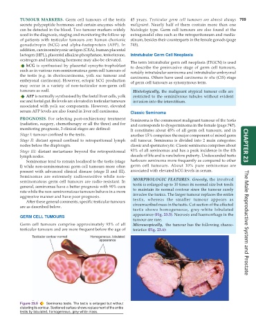
seminomatous germ cell tumours are radio-resistant. In MORPHOLOGIC FEATURES. Grossly, the involved
general, seminomas have a better prognosis with 90% cure testis is enlarged up to 10 times its normal size but tends
rate while the non-seminomatous tumours behave in a more to maintain its normal contour since the tumour rarely
aggressive manner and have poor prognosis. invades the tunica. The larger tumour replaces the entire
After these general comments, specific testicular tumours testis, whereas the smaller tumour appears as
are as described below. circumscribed mass in the testis. Cut section of the affected
testis shows homogeneous, grey-white lobulated
appearance (Fig. 23.5). Necrosis and haemorrhage in the
GERM CELL TUMOURS
tumour are rare.
Germ cell tumours comprise approximately 95% of all Microscopically, the tumour has the following charac- The Male Reproductive System and Prostate
testicular tumours and are more frequent before the age of teristics (Fig. 23.6):
Figure 23.5 Seminoma testis. The testis is enlarged but without
distorting its contour. Sectioned surface shows replacement of the entire
testis by lobulated, homogeneous, grey-white mass.