
Page 727 - Textbook of Pathology, 6th Edition
P. 727
tumour giant cells are frequently present. Haemorrhage 4. There may be presence of both intracellular and 711
and necrosis are common. extracellular PAS-positive hyaline globules, many of which
3. The stroma is not as distinct as in seminoma and may contain AFP.
contain variable amount of primitive mesenchyme.
Polyembryoma
Embryonal carcinoma is more aggressive and less
radiosensitive than seminoma. Chemotherapy is more Polyembryoma is defined as a tumour composed predo-
effective in treating this tumour. minantly of embryoid bodies. Embryoid bodies are structures
containing a disc and cavities surrounded by loose
Yolk Sac Tumour mesenchyme simulating an embryo of about 2 weeks’
(Synonyms: Endodermal Sinus Tumour, gestation. Polyembryoma is extremely rare but embryoid
Orchioblastoma, Infantile Embryonal Carcinoma) bodies may be present with embryonal carcinoma and
teratoma.
This characteristic tumour is the most common testicular
tumour of infants and young children upto the age of 4 years. Choriocarcinoma
In adults, however, yolk sac tumour in pure form is rare but
may be present as the major component in 40% of germ cell Pure choriocarcinoma is a highly malignant tumour compo-
tumours. AFP levels are elevated in 100% cases of yolk sac sed of elements consisting of syncytiotrophoblast and
tumours. cytotrophoblast.
However, pure form is extremely rare and occurs more
MORPHOLOGIC FEATURES. Grossly, the tumour is often in combination with other germ cell tumours. The
generally soft, yellow-white, mucoid with areas of necrosis patients are generally in their 2nd decade of life. The primary
and haemorrhages. tumour is usually small and the patient may manifest initially
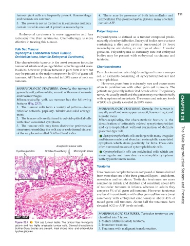
Microscopically, yolk sac tumour has the following with symptoms of metastasis. The serum and urinary levels
features (Fig. 23.7): of hCG are greatly elevated in 100% cases.
1. The tumour cells form a variety of patterns—loose MORPHOLOGIC FEATURES. Grossly, the tumour is CHAPTER 23
reticular network, papillary, tubular and solid arrange- usually small and may appear as a soft, haemorrhagic and
ment. necrotic mass.
2. The tumour cells are flattened to cuboid epithelial cells Microscopically, the characteristic feature is the
with clear vacuolated cytoplasm. identification of intimately related syncytiotrophoblast
3. The tumour cells may form distinctive perivascular and cytotrophoblast without formation of definite
structures resembling the yolk sac or endodermal sinuses placental-type villi.
of the rat placenta called Schiller-Duval bodies.
Syncytiotrophoblastic cells are large with many irregular
and bizarre nuclei and abundant eosinophilic vacuolated
cytoplasm which stains positively for hCG. These cells
often surround masses of cytotrophoblastic cells.
Cytotrophoblastic cells are polyhedral cells which are
more regular and have clear or eosinophilic cytoplasm
with hyperchromatic nuclei.
Teratoma The Male Reproductive System and Prostate
Teratomas are complex tumours composed of tissues derived
from more than one of the three germ cell layers—endoderm,
mesoderm and ectoderm. Testicular teratomas are more
common in infants and children and constitute about 40%
of testicular tumours in infants, whereas in adults they
comprise 5% of all germ cell tumours. However, teratomas
are found in combination with other germ cell tumours (most
commonly with embryonal carcinoma) in about 45% of
mixed germ cell tumours. About half the teratomas have
elevated hCG or AFP levels or both.
MORPHOLOGIC FEATURES. Testicular teratomas are
classified into 3 types:
1. Mature (differentiated) teratoma
Figure 23.7 Yolk sac tumour testis. The tumour has microcystic
pattern and has highly anaplastic tumour cells. Several characteristic 2. Immature teratoma
Schiller-Duval bodies are present. Inset shows intra- and extracellular 3. Teratoma with malignant transformation.
hyaline globules.