
Page 728 - Textbook of Pathology, 6th Edition
P. 728
712
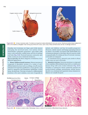
Figure 23.8 Immature teratoma testis. The testis is enlarged and nodular distorting the testicular contour. Sectioned surface shows replacement
of the entire testis by variegated mass having grey-white solid areas, cystic areas, honey-combed areas and foci of cartilage and bone.
Grossly, most teratomas are large, grey-white masses infants and children and has favourable prognosis.
enlarging the involved testis. Cut surface shows However, similar mature and benign-appearing tumour
characteristic variegated appearance—grey-white solid in adults is invariably associated with small hidden foci
areas, cystic and honey-combed areas, and foci of cartilage of immature elements so that their clinical course in adults
and bone (Fig. 23.8). Dermoid tumours commonly seen is unpredictable. It is believed that all testicular teratomas
SECTION III
in the ovaries are rare in testicular teratomas. in the adults are malignant.
Microscopically, the three categories of teratomas show As mentioned above, dermoid cysts similar to those
different appearances: of the ovary are rare in the testis.
1. Mature (differentiated) teratoma. Mature teratoma is 2. Immature teratoma. Immature teratoma is composed
composed of disorderly mixture of a variety of well- of incompletely differentiated and primitive or embryonic
differentiated structures such as cartilage, smooth muscle, tissues along with some mature elements (Fig. 23.9).
intestinal and respiratory epithelium, mucus glands, cysts Primitive or embryonic tissue commonly present are
lined by squamous and transitional epithelium, neural poorly-formed cartilage, mesenchyme, neural tissues,
tissue, fat and bone. This type of mature or differentiated abortive eye, intestinal and respiratory tissue elements etc.
teratoma is the most common, seen more frequently in Mitoses are usually frequent.
Systemic Pathology
Figure 23.9 Immature teratoma testis. Microscopy shows a variety of incompletely differentiated tissue elements.