
Page 726 - Textbook of Pathology, 6th Edition
P. 726
710
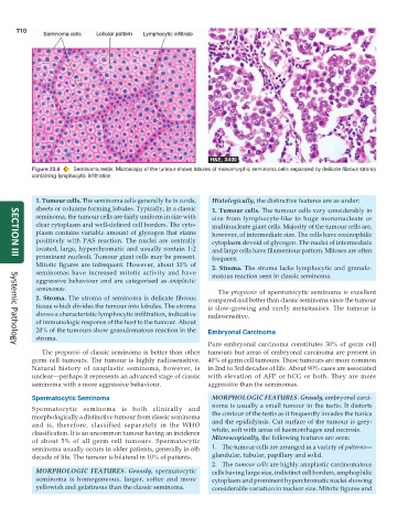
Figure 23.6 Seminoma testis. Microscopy of the tumour shows lobules of monomorphic seminoma cells separated by delicate fibrous stroma
containing lymphocytic infiltration.
1. Tumour cells. The seminoma cells generally lie in cords, Histologically, the distinctive features are as under:
sheets or columns forming lobules. Typically, in a classic 1. Tumour cells. The tumour cells vary considerably in
seminoma, the tumour cells are fairly uniform in size with size from lymphocyte-like to huge mononucleate or
clear cytoplasm and well-defined cell borders. The cyto- multinucleate giant cells. Majority of the tumour cells are,
plasm contains variable amount of glycogen that stains however, of intermediate size. The cells have eosinophilic
positively with PAS reaction. The nuclei are centrally cytoplasm devoid of glycogen. The nuclei of intermediate
located, large, hyperchromatic and usually contain 1-2 and large cells have filamentous pattern. Mitoses are often
prominent nucleoli. Tumour giant cells may be present. frequent.
SECTION III
Mitotic figures are infrequent. However, about 10% of 2. Stroma. The stroma lacks lymphocytic and granulo-
seminomas have increased mitotic activity and have matous reaction seen in classic seminoma.
aggressive behaviour and are categorised as anaplastic
seminomas. The prognosis of spermatocytic seminoma is excellent
2. Stroma. The stroma of seminoma is delicate fibrous compared and better than classic seminoma since the tumour
tissue which divides the tumour into lobules. The stroma is slow-growing and rarely metastasises. The tumour is
shows a characteristic lymphocytic infiltration, indicative radiosensitive.
of immunologic response of the host to the tumour. About
20% of the tumours show granulomatous reaction in the Embryonal Carcinoma
stroma.
Pure embryonal carcinoma constitutes 30% of germ cell
Systemic Pathology
The prognosis of classic seminoma is better than other tumours but areas of embryonal carcinoma are present in
germ cell tumours. The tumour is highly radiosensitive. 40% of germ cell tumours. These tumours are more common
Natural history of anaplastic seminoma, however, is in 2nd to 3rd decades of life. About 90% cases are associated
unclear—perhaps it represents an advanced stage of classic with elevation of AFP or hCG or both. They are more
seminoma with a more aggressive behaviour. aggressive than the seminomas.
Spermatocytic Seminoma MORPHOLOGIC FEATURES. Grossly, embryonal carci-
noma is usually a small tumour in the testis. It distorts
Spermatocytic seminoma is both clinically and
morphologically a distinctive tumour from classic seminoma the contour of the testis as it frequently invades the tunica
and is, therefore, classified separately in the WHO and the epididymis. Cut surface of the tumour is grey-
classification. It is an uncommon tumour having an incidence white, soft with areas of haemorrhages and necrosis.
of about 5% of all germ cell tumours. Spermatocytic Microscopically, the following features are seen:
seminoma usually occurs in older patients, generally in 6th 1. The tumour cells are arranged in a variety of patterns—
decade of life. The tumour is bilateral in 10% of patients. glandular, tubular, papillary and solid.
2. The tumour cells are highly anaplastic carcinomatous
MORPHOLOGIC FEATURES. Grossly, spermatocytic cells having large size, indistinct cell borders, amphophilic
seminoma is homogeneous, larger, softer and more cytoplasm and prominent hyperchromatic nuclei showing
yellowish and gelatinous than the classic seminoma. considerable variation in nuclear size. Mitotic figures and