
Page 761 - Textbook of Pathology, 6th Edition
P. 761
tiating towards endometrial type of epithelium. In view of carcinoma i.e. clear cells having abundant eosinophilic 745
presence of endometriosis in a few cases of endometrioid cytoplasm rich in glycogen.
tumours, some authors have suggested malignant trans-
formation of endometriosis. Brenner Tumour
MORPHOLOGIC FEATURES. Grossly, these tumours Brenner tumours are uncommon and comprise about 2% of
are partly solid and partly cystic and may have foci of all ovarian tumours. They are characteristically solid ovarian
haemorrhages, especially in benign variety. tumours. Less than 10% of Brenner tumours are bilateral.
Histologically, the endometrioid adenocarcinoma is Most Brenner tumours are benign. Rarely, borderline form
distinguished from serous and mucinous carcinomas by is encountered called ‘proliferating Brenner tumour’ while
typical glandular pattern that closely resembles that of the one with carcinomatous change is termed ‘malignant
uterine endometrioid adenocarcinoma. There may be foci Brenner tumour’.
of squamous metaplasia justifying the diagnosis of Histogenesis of the tumour is from coelomic epithelium
adenoacanthoma. Papillary pattern and foci of serous and by metaplastic transformation into transitional epithelium
mucinous carcinoma may also be found. Benign variety (urothelium).
closely resembles endometriosis with cystic change. There
are no clearly defined criteria for borderline endometrioid MORPHOLOGIC FEATURES. Grossly, Brenner tumour
tumours. is typically solid, yellow-grey, firm mass of variable size.
Occasionally, a few scattered tiny cysts may be present
Clear Cell (Mesonephroid) Tumours on cut section.
Clear cell (mesonephroid) tumours are almost always Histologically, Brenner tumour consists of nests, masses
malignant and comprise about 5% of all ovarian cancers; rare and columns of epithelial cells, scattered in fibrous stroma
benign variety is called clear cell adenofibroma. They are termed of the ovary. These epithelial cells resemble urothelial cells
clear cell or mesonephroid carcinomas because of the close which are ovoid in shape, having clear cytoplasm,
histologic resemblance to renal adenocarcinoma. They have vesicular nuclei with characteristic nuclear groove called
also been called as mesonephroma or mesonephric carcinoma ‘coffee-bean’ nuclei.
because of the questionable relationship to the mesonephric CHAPTER 24
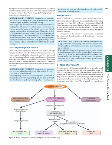
structures. II. GERM CELL TUMOURS
Ovarian germ cell tumours arising from germ cells which
MORPHOLOGIC FEATURES. Grossly, these tumours produce the female gametes (i.e. ova) account for about 15-
are large, usually unilateral, partly solid and partly cystic. 20% of all ovarian neoplasms. The neoplastic germ cells may
Less than 10% are bilateral. follow one of the several lines of differentiation as shown in
Histologically, clear cell or mesonephroid carcinoma is Fig. 24.25. Nearly 95% of them are benign and occur chiefly
characterised by tubules, glands, papillae, cysts and solid
sheets of tumour cells resembling cells of renal adeno- in young females, vast majority of them being benign cystic
teratomas (dermoid cysts). The remainder are malignant The Female Genital Tract
Figure 24.25 Histogenetic classification of germ cell tumours of the ovary.