
Page 762 - Textbook of Pathology, 6th Edition
P. 762
746
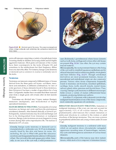
Figure 24.26 Dermoid cyst of the ovary. The ovary is enlarged and
shows a large unilocular cyst containing hair, pultaceous material and
bony tissue.
germ cell tumours comprising a variety of morphologic forms seen (Rokitansky’s protuberance) where tissue elements
occurring chiefly in children and young adults and are highly such as tooth, bone, cartilage and various other odd tissues
aggressive tumours. Most germ cell tumours of the ovaries are present (Fig. 24.26). Less often, the cyst may contain
have their counterparts in the testis (Chapter 23) and mucoid material.
sometimes in the mediastinum but their frequency differs Microscopically, the most prominent feature is the lining
from one site to the other. For instance, benign cystic teratoma of the cyst wall by stratified squamous epithelium and its
or dermoid cyst so common in ovaries is extremely rare in adnexal structures such as sebaceous glands, sweat glands
SECTION III
the testis. and hair follicles (Fig. 24.27). Though ectodermal
derivatives are most prominent features, tissues of
Teratomas
mesodermal and endodermal origin are also commonly
Teratomas are tumours composed of different types of tissues present. Various other tissue components frequently
derived from the three germ cell layers—ectoderm, meso- found in teratomas are bronchus, intestinal epithelium,
derm and endoderm, in different combinations. In view of cartilage, bone, tooth, smooth muscle, neural tissue,
wide spectrum of tissue elements found in these teratomas, salivary gland, retina, pancreas and thyroid tissue. Thus,
their histogenesis has been a matter of speculation for a long viewing a benign cystic teratoma in different microscopic
time. Cytogenetic studies have revealed that these tumours fields reveals a variety of mature differentiated tissue
arise from a single germ cell (ovum) after its first meiotic elements, producing kaleidoscopic patterns.
division. Less than 1% of patients with a dermoid cyst develop
Systemic Pathology
Teratomas are divided into 3 types: mature (benign), malignant transformation of one of the tissue components,
immature (malignant), and monodermal or highly most commonly squamous cell carcinoma.
specialised teratomas.
IMMATURE (MALIGNANT) TERATOMA. Immature or
MATURE (BENIGN) TERATOMA. Vast majority of ovarian malignant teratomas of the ovary are rare and account for
teratomas are benign and cystic and have the predominant approximately 0.2% of all ovarian tumours. They are
ectodermal elements, often termed clinically as dermoid cyst. predominantly solid tumours that contain immature or
Infrequently, mature teratoma may be solid and benign and embryonal structures in contrast to the mature or adult
has to be distinguished from immature or malignant structures of the benign teratomas. They are more common
teratoma. Benign cystic teratomas are more frequent in young in prepubertal adolescents and young women under 20 years
women during their active reproductive life. The tumour is of age.
bilateral in 10% of cases.
Grossly, malignant teratoma is a unilateral solid mass
Grossly, benign cystic teratoma or dermoid cyst is
characteristically a unilocular cyst, 10-15 cm in diameter, which on cut section shows characteristic variegated
usually lined by the skin and hence its name. On appearance revealing areas of haemorrhages, necrosis,
sectioning, the cyst is filled with paste-like sebaceous tiny cysts and heterogeneous admixture of various tissue
secretions and desquamated keratin admixed with masses elements.
of hair. The cyst wall is thin and opaque grey-white. Gene- Microscopically, parts of the tumour may show mature
rally, in one area of the cyst wall, a solid prominence is tissues, while most of it is composed of immature tissues