


Page 78 - Textbook of Pathology, 6th Edition
P. 78
62
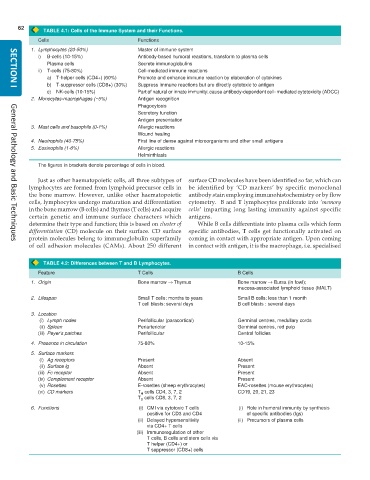
TABLE 4.1: Cells of the Immune System and their Functions.
Cells Functions
1. Lymphocytes (20-50%) Master of immune system
i) B-cells (10-15%) Antibody-based humoral reactions, transform to plasma cells
Plasma cells Secrete immunoglobulins
ii) T-cells (75-80%) Cell-mediated immune reactions
a) T-helper cells (CD4+) (60%) Promote and enhance immune reaction by elaboration of cytokines
b) T-suppressor cells (CD8+) (30%) Suppress immune reactions but are directly cytotoxic to antigen
SECTION I
c) NK-cells (10-15%) Part of natural or innate immunity; cause antibody-dependent cell- mediated cytotoxicity (ADCC)
2. Monocytes-macrophages (~5%) Antigen recognition
Phagocytosis
Secretory function
Antigen presentation
3. Mast cells and basophils (0-1%) Allergic reactions
Wound healing
4. Neutrophils (40-75%) First line of dense against microorganisms and other small antigens
5. Eosinophils (1-6%) Allergic reactions
Helminthiasis
The figures in brackets denote percentage of cells in blood.
Just as other haematopoietic cells, all three subtypes of surface CD molecules have been identified so far, which can
lymphocytes are formed from lymphoid precursor cells in be identified by ‘CD markers’ by specific monoclonal
the bone marrow. However, unlike other haematopoietic antibody stain employing immunohistochemistry or by flow
cells, lymphocytes undergo maturation and differentiation cytometry. B and T lymphocytes proliferate into ‘memory
in the bone marrow (B cells) and thymus (T cells) and acquire cells’ imparting long lasting immunity against specific
certain genetic and immune surface characters which antigens.
determine their type and function; this is based on cluster of While B cells differentiate into plasma cells which form
differentiation (CD) molecule on their surface. CD surface specific antibodies, T cells get functionally activated on
protein molecules belong to immunoglobulin superfamily coming in contact with appropriate antigen. Upon coming
General Pathology and Basic Techniques
of cell adhesion molecules (CAMs). About 250 different in contact with antigen, it is the macrophage, i.e. specialised
TABLE 4.2: Differences between T and B Lymphocytes.
Feature T Cells B Cells
1. Origin Bone marrow → Thymus Bone marrow → Bursa (in fowl);
mucosa-associated lymphoid tissue (MALT)
2. Lifespan Small T cells: months to years Small B cells: less than 1 month
T cell blasts: several days B cell blasts : several days
3. Location
(i) Lymph nodes Perifollicular (paracortical) Germinal centres, medullary cords
(ii) Spleen Periarteriolar Germinal centres, red pulp
(iii) Peyer’s patches Perifollicular Central follicles
4. Presence in circulation 75-80% 10-15%
5. Surface markers
(i) Ag receptors Present Absent
(ii) Surface lg Absent Present
(iii) Fc receptor Absent Present
(iv) Complement receptor Absent Present
(v) Rosettes E-rosettes (sheep erythrocytes) EAC-rosettes (mouse erythrocytes)
(vi) CD markers T cells CD4, 3, 7, 2 CD19, 20, 21, 23
H
T cells CD8, 3, 7, 2
S
6. Functions (i) CMI via cytotoxic T cells (i) Role in humoral immunity by synthesis
positive for CD3 and CD4 of specific antibodies (Igs)
(ii) Delayed hypersensitivity (ii) Precursors of plasma cells
via CD4+ T cells
(iii) Immunoregulation of other
T cells, B cells and stem cells via
T helper (CD4+) or
T suppressor (CD8+) cells

