


Page 74 - Textbook of Pathology, 6th Edition
P. 74
58


     SECTION I


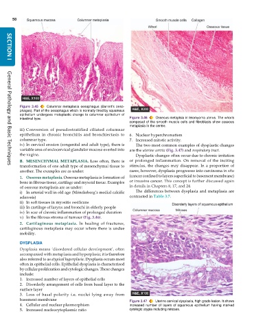
           Figure 3.45  Columnar metaplasia oesophagus (Barrett’s oeso-
           phagus). Part of the oesophagus which is normally lined by squamous
           epithelium undergoes metaplastic change to columnar epithelium of
           intestinal type.                                    Figure 3.46  Osseous metaplsia in leiomyoma uterus. The whorls
                                                               composed of the smooth muscle cells and fibroblasts show osseous
                                                               metaplasia in the centre.
           iii) Conversion of pseudostratified ciliated columnar
           epithelium in chronic bronchitis and bronchiectasis to  6. Nuclear hyperchromatism
           columnar type.                                      7. Increased mitotic activity.
           iv) In cervical erosion (congenital and adult type), there is  The two most common examples of dysplastic changes
           variable area of endocervical glandular mucosa everted into  are the uterine cervix (Fig. 3.47) and respiratory tract.
           the vagina.                                            Dysplastic changes often occur due to chronic irritation
           B. MESENCHYMAL METAPLASIA. Less often, there is     or prolonged inflammation. On removal of the inciting
           transformation of one adult type of mesenchymal tissue to  stimulus, the changes may disappear. In a proportion of
     General Pathology and Basic Techniques
           another. The examples are as under:                 cases, however, dysplasia progresses into carcinoma in situ
           1. Osseous metaplasia. Osseous metaplasia is formation of  (cancer confined to layers superficial to basement membrane)
           bone in fibrous tissue, cartilage and myxoid tissue. Examples  or invasive cancer. This concept is further discussed again
           of osseous metaplasia are as under:                 in details in Chapters 8, 17, and 24.
           i) In arterial wall in old age (Mönckeberg’s medial calcific  The differences between dysplasia and metaplasia are
           sclerosis)                                          contrasted in Table 3.7.
           ii) In soft tissues in myositis ossificans
           iii) In cartilage of larynx and bronchi in elderly people
           iv) In scar of chronic inflammation of prolonged duration
           v) In the fibrous stroma of tumour (Fig. 3.46).
           2. Cartilaginous  metaplasia.  In healing of fractures,
           cartilaginous metaplasia may occur where there is undue
           mobility.
           DYSPLASIA
           Dysplasia means ‘disordered cellular development’, often
           accompanied with metaplasia and hyperplasia; it is therefore
           also referred to as atypical hyperplasia. Dysplasia occurs most
           often in epithelial cells. Epithelial dysplasia is characterised
           by cellular proliferation and cytologic changes. These changes
           include:
           1. Increased number of layers of epithelial cells
           2. Disorderly arrangement of cells from basal layer to the
           surface layer
           3. Loss of basal polarity i.e. nuclei lying away from
           basement membrane                                   Figure 3.47  Uterine cervical dysplasia, high grade lesion. It shows
           4. Cellular and nuclear pleomorphism                increased number of layers of squamous epithelium having marked
           5. Increased nucleocytoplasmic ratio                cytologic atypia including mitoses.

