
Page 799 - Textbook of Pathology, 6th Edition
P. 799
ii) Pronounced parakeratosis. Cancer of scrotal skin in chimney-sweeps was the first 783
iii) Marked epidermal hyperplasia with disappearance of cancer in which an occupational carcinogen (soot) was impli-
dermal papillae. cated. ‘Kangari cancer’ of the skin of inner side of thigh and
iv) Scattered bizarre dyskeratotic cells distributed lower abdomen common in natives of Kashmir is another
throughout the epidermis. example of skin cancer due to chronic irritation (Kangari is
an earthenware pot containing glowing charcoal used by
Bowen’s disease, unlike solar keratosis which invariably Kashmiris close to their abdomen to keep them warm).
leads to invasive cancer, may remain confined to the surface Although squamous carcinomas can occur anywhere on
for many years. the skin, most common locations are the face, pinna of the
3. XERODERMA PIGMENTOSUM. This condition is a ears, back of hands and mucocutaneous junctions such as
hypersensitivity of the skin to sunlight that is determined on the lips, anal canal and glans penis. Cutaneous squamous
by a recessive gene. The disorder may lead to multiple carcinoma arising in a pre-existing inflammatory and
malignancies of the skin such as basal cell carcinoma, degenerative lesion has a higher incidence of developing
squamous cell carcinoma and malignant melanoma. metastases.
Xeroderma pigmentosum has already been described under MORPHOLOGIC FEATURES. Grossly, squamous
genetic dermatoses. carcinoma of the skin and squamous-lined mucosa can
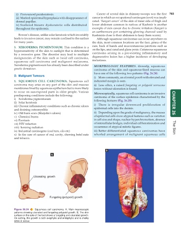
have one of the following two patterns (Fig. 26.24):
D. Malignant Tumours
i) More commonly, an ulcerated growth with elevated and
1. SQUAMOUS CELL CARCINOMA. Squamous cell indurated margin is seen.
carcinoma may arise on any part of the skin and mucous ii) Less often, a raised fungating or polypoid verrucous
membranes lined by squamous epithelium but is more likely lesion without ulceration is found.
to occur on sun-exposed parts in older people. Various Microscopically, squamous cell carcinoma is an invasive
predisposing conditions include the following: carcinoma of the surface epidermis characterised by the
i) Xeroderma pigmentosum following features (Fig. 26.25):
ii) Solar keratosis CHAPTER 26
iii) Chronic inflammatory conditions such as chronic ulcers i) There is irregular downward proliferation of
and draining osteomyelitis epidermal cells into the dermis.
iv) Old burn scars (Marjolin’s ulcers) ii) Depending upon the grade of malignancy, the masses
v) Chemical burns of epidermal cells show atypical features such as variation
vi) Psoriasis in cell size and shape, nuclear hyperchromatism, absence
vii) HIV infection of intercellular bridges, individual cell keratinisation and
viii) Ionising radiation occurrence of atypical mitotic figures. The Skin
ix) Industrial carcinogens (coal tars, oils etc) iii) Better-differentiated squamous carcinomas have
x) In the case of cancer of oral cavity, chewing betel nuts whorled arrangement of malignant squamous cells
and tobacco.
Figure 26.24 Squamous cell carcinoma. A, Main macroscopic
patterns showing ulcerated and fungating polypoid growth. B, The skin
surface on the sole of the foot shows a fungating and ulcerated growth.
On cutting, the growth is both exophytic and endophytic and is chalky
white in colour.