
Page 858 - Textbook of Pathology, 6th Edition
P. 858
842
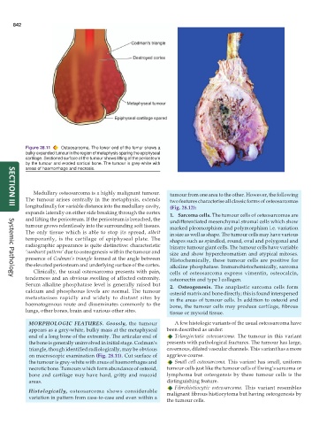
Figure 28.11 Osteosarcoma. The lower end of the femur shows a
bulky expanded tumour in the region of metaphysis sparing the epiphyseal
cartilage. Sectioned surface of the tumour shows lifting of the periosteum
by the tumour and eroded cortical bone. The tumour is grey-white with
areas of haemorrhage and necrosis.
Medullary osteosarcoma is a highly malignant tumour. tumour from one area to the other. However, the following
The tumour arises centrally in the metaphysis, extends two features characterise all classic forms of osteosarcomas
longitudinally for variable distance into the medullary cavity, (Fig. 28.12):
SECTION III
expands laterally on either side breaking through the cortex 1. Sarcoma cells. The tumour cells of osteosarcomas are
and lifting the periosteum. If the periosteum is breached, the undifferentiated mesenchymal stromal cells which show
tumour grows relentlessly into the surrounding soft tissues. marked pleomorphism and polymorphism i.e. variation
The only tissue which is able to stop its spread, albeit in size as well as shape. The tumour cells may have various
temporarily, is the cartilage of epiphyseal plate. The shapes such as spindled, round, oval and polygonal and
radiographic appearance is quite distinctive: characteristic bizarre tumour giant cells. The tumour cells have variable
‘sunburst pattern’ due to osteogenesis within the tumour and size and show hyperchromatism and atypical mitoses.
presence of Codman’s triangle formed at the angle between Histochemically, these tumour cells are positive for
the elevated periosteum and underlying surface of the cortex. alkaline phosphatase. Immunohistochemically, sarcoma
Clinically, the usual osteosarcoma presents with pain, cells of osteosarcoma express vimentin, osteocalcin,
tenderness and an obvious swelling of affected extremity. osteonectin and type I collagen.
Systemic Pathology
Serum alkaline phosphatase level is generally raised but 2. Osteogenesis. The anaplastic sarcoma cells form
calcium and phosphorus levels are normal. The tumour osteoid matrix and bone directly; this is found interspersed
metastasises rapidly and widely to distant sites by in the areas of tumour cells. In addition to osteoid and
haematogenous route and disseminates commonly to the bone, the tumour cells may produce cartilage, fibrous
lungs, other bones, brain and various other sites. tissue or myxoid tissue.
MORPHOLOGIC FEATURES. Grossly, the tumour A few histologic variants of the usual osteosarcoma have
appears as a grey-white, bulky mass at the metaphyseal been described as under:
end of a long bone of the extremity. The articular end of Telangiectatic osteosarcoma. The tumour in this variant
the bone is generally uninvolved in initial stage. Codman’s presents with pathological fractures. The tumour has large,
triangle, though identified radiologically, may be obvious cavernous, dilated vascular channels. This variant has a more
on macroscopic examination (Fig. 28.11). Cut surface of aggrieve course.
the tumour is grey-white with areas of haemorrhages and Small cell osteosarcoma. This variant has small, uniform
necrotic bone. Tumours which form abundance of osteoid, tumour cells just like the tumour cells of Ewing’s sarcoma or
bone and cartilage may have hard, gritty and mucoid lymphoma but osteogensis by these tumour cells is the
areas. distinguishing feature.
Fibrohistiocytic osteosarcoma. This variant resembles
Histologically, osteosarcoma shows considerable malignant fibrous histiocytoma but having osteogenesis by
variation in pattern from case-to-case and even within a
the tumour cells.