
Page 859 - Textbook of Pathology, 6th Edition
P. 859
843
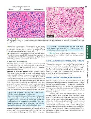
Figure 28.12 Osteosarcoma. Hallmarks of microscopic picture of the usual osteosarcoma are the sarcoma cells characterised by variation in
size and shape of tumour cells, bizarre mitosis and multinucleate tumour giant cells, and osteogenesis i.e. production of osteoid matrix and bone
directly by the tumour cells.
Anaplastic osteosarcoma. In this variant, the tumour has so Microscopically, periosteal osteosarcoma has cartilaginous
marked anaplasia that it may resemble any other type of differentiation and higher degree of anaplasia than that
pleomorphic sarcoma and is identified by the presence of seen in parosteal osteosarcoma.
osteoid formed directly by the tumour cells.
Well-differentiated osteosarcoma. Although generally classic Table 28.3 sums up the contrasting features of central CHAPTER 28
form of osteosarcoma is a highly malignant tumour, rarely a (medullary) and surface (parosteal and periosteal)
well-differentiated variant having minimal cytologic atypia osteosarcomas.
resembling parosteal osteosarcoma may be seen.
SURFACE OSTEOSARCOMA CARTILAGE-FORMING (CHONDROBLASTIC) TUMOURS
About 5% of osteosarcomas occur on the surface of bone and The tumours which are composed of frank cartilage or
are slow-growing tumours compared to medullary derived from cartilage-forming cells are included in this
osteosarcomas. Surface osteosarcoma includes 2 variants: group. This group comprises benign lesions like osteocartila-
parosteal and periosteal. ginous exostoses (osteochondromas), enchondroma,
Parosteal or juxtacortical osteosarcoma is an uncommon chondroblastoma and chondromyxoid fibroma, and a
form of osteosarcoma having its origin from the metaphysis malignant counterpart, chondrosarcoma. The Musculoskeletal System
on the external surface of the bone (parosteal or juxtacortical
means outer to cortex). The tumour should be distinguished Osteocartilaginous Exostoses (Osteochondromas)
from the more common medullary osteosarcoma because of
its better prognosis and different presentation. The tumour Osteocartilaginous exostoses or osteochondromas are the
occurs in older age group, has no sex predilection and is slow commonest of benign cartilage-forming lesions. Though
growing. Its common locations are metaphysis of long bones, designated and discussed with neoplasms, exostosis or
most frequently lower end of the femur and upper end of osteochondroma is not a true tumour but is regarded as a
the humerus. X-ray examination usually reveals a dense bony disorder of growth and development (page 834). It may occur
mass attached to the outer cortex of the affected long bone. as a ‘solitary sporadic exostosis’ or there may be ‘multiple
Grossly, the tumour is lobulated and circumscribed, hereditary exostoses’.
calcified mass in the subperiosteal location. Exostoses arise from metaphyses of long bones as
Microscopically, the features which characterise the usual exophytic lesions, most commonly lower femur and upper
osteosarcoma (sarcomatous stroma and production of tibia (i.e. around knee) and upper humerus but may also be
neoplastic osteoid and bone) are present, but the tumour found in other bones such as the scapula or ilium. They are
shows a high degree of structural differentiation, and there discovered most commonly in late childhood or adolescence
are generally well-formed bony trabeculae. These features and are more frequent in males. They may remain
account for distinctly better prognosis in these cases. asymptomatic and discovered as an incidental radiographic
finding or may produce obvious deformity. Both solitary and
Periosteal osteosarcoma is a rare form of osteosarcoma that multiple exostoses may undergo transformation into
arises between the cortex and the overlying periosteum. Its chondrosarcoma but the risk is much greater with multiple
common location is the diaphysis of the tibia or the femur. hereditary exostoses.