
Page 860 - Textbook of Pathology, 6th Edition
P. 860
844
TABLE 28.3. Contrasting Features of Central (Medullary) and Surface (Parosteal and Periosteal) Osteosarcoma.
Feature Central (Medullary) Surface (Parosteal and Periosteal)
1. Age 10-20 years Older patients
2. Sex More common in males No sex predilection
3. Anatomic site Metaphysis Metaphysis/diaphysis
4. Location Femur (lower end), tibia (upper end), Femur (lower-end), humerus (upper end)
humerus (upper end), around hip
5. Pathogenesis Primary: genetic factors Parosteal: Arises outer to cortex
(mutations in Rb gene, p53, MDM2) Periosteal: Arises between cortex and
Secondary: Paget’s disease, fibrous dysplasia periosteum
6. Behaviour Highly malignant Slow growing
7. G/A Bulky, necrotic, forms Codman’s triangle Smaller, well-formed bone present
8. M/E i. Sarcomas cells: Polymorphic and pleomorphic i. Parosteal: Fibrous stromal cells with
ii. Osteoid formation subtle atypia
ii. Periosteal: High grade
iii. Both form bony trabeculae
9. Histologic types Telangiectatic, small cell, fibrohistiocystic, Parosteal (juxta cortical), periosteal
well differentiated, anaplastic
10. Spread and prognosis Haematogenous spread, Recurrences common, may metastasise,
prognosis poor prognosis generally good, better for
parosteal than periosteal
Enchondroma
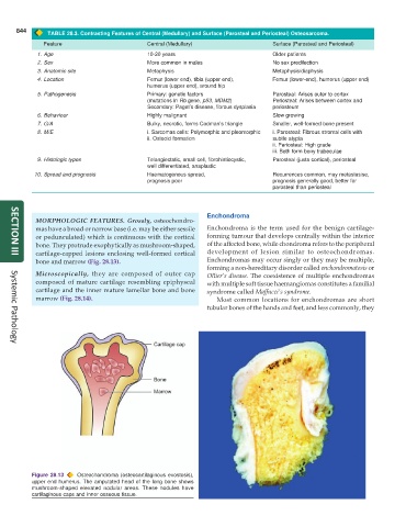
MORPHOLOGIC FEATURES. Grossly, osteochondro-
mas have a broad or narrow base (i.e. may be either sessile Enchondroma is the term used for the benign cartilage-
or pedunculated) which is continuous with the cortical forming tumour that develops centrally within the interior
bone. They protrude exophytically as mushroom-shaped, of the affected bone, while chondroma refers to the peripheral
cartilage-capped lesions enclosing well-formed cortical development of lesion similar to osteochondromas.
SECTION III
bone and marrow (Fig. 28.13). Enchondromas may occur singly or they may be multiple,
forming a non-hereditary disorder called enchondromatosis or
Microscopically, they are composed of outer cap Ollier’s disease. The coexistence of multiple enchondromas
composed of mature cartilage resembling epiphyseal with multiple soft tissue haemangiomas constitutes a familial
cartilage and the inner mature lamellar bone and bone syndrome called Maffucci’s syndrome.
marrow (Fig. 28.14). Most common locations for enchondromas are short
tubular bones of the hands and feet, and less commonly, they
Systemic Pathology
Figure 28.13 Osteochondroma (osteocartilaginous exostosis),
upper end humerus. The amputated head of the long bone shows
mushroom-shaped elevated nodular areas. These nodules have
cartilaginous caps and inner osseous tissue.