
Page 878 - Textbook of Pathology, 6th Edition
P. 878
862 KELOID. A keloid is a progressive fibrous overgrowth in
response to cutaneous injury such as burns, incisions, insect
bites, vaccinations and others. Keloids are found more often
in blacks. Their excision is frequently followed by
recurrences.
Grossly, the keloid is a firm, smooth, pink, raised patch
from which extend claw-like processes (keloid-claw).
Histologically, it is composed of thick, homogeneous,
eosinophilic hyalinised bands of collagen admixed with
thin collagenous fibres and large active fibroblasts. The
adnexal structures are atrophic or destroyed.
There are some differences between a keloid and a
hypertrophic scar. A hypertrophic scar of the skin is more
cellular and has numerous fibroblasts than a keloid and is
composed of thinner collagenous fibres. A keloid is a
progressive lesion and liable to recurrences after surgical
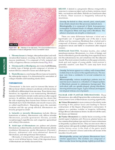
Figure 29.1 Fibroma of the oral cavity. The circumscribed lesion is excision.
composed of mature collagenised fibrous connective tissue.
NODULAR FASCITIS. Nodular fascitis, also called
pseudosarcomatous fibromatosis, is a form of benign and
1. Fibroma durum is a benign, often pedunculated and well- reactive fibroblastic growth extending from superficial fascia
circumscribed tumour occurring on the body surfaces and into the subcutaneous fat, and sometimes into the subjacent
mucous membranes. It is composed of fully matured and muscle. The most common locations are the upper extremity,
richly collagenous fibrous connective tissue (Fig. 29.1). trunk and neck region of young adults. Local excision is
generally curative. Less than 5% cases may have local
2. Fibroma molle or fibrolipoma, also termed soft fibroma, recurrence.
is similar type of benign growth composed of mixture of
mature fibrous connective tissue and adult-type fat. Grossly, the lesion appears as a solitary well-cirumscribed
nodule (true to its name) in the superficial fascia. The size
SECTION III
3. Elastofibroma is a rare benign fibrous tumour located in may vary from a centimeter to several centimeters in
the subscapular region. It is characterised by association of diameter.
collagen bundles and branching elastic fibres.
Microscopically, various morphologic patterns may be
seen but most common is a whorled or S-shaped pattern
FIBROMATOSIS
of fibroblasts present in oedematous background. The
‘Fibromatosis’ is the term used for tumour-like lesions of individual cells are spindle-shaped, plump fibroblasts
fibrous tissue which continue to proliferate actively and may showing mild nuclear atypia. Typical mitoses are frequent
be difficult to differentiate from sarcomas. These lesions may, but atypical mitoses are not present.
therefore, be regarded as non-metastasising fibroblastic
tumours which tend to invade locally and recur after surgical PALMAR AND PLANTAR FIBROMATOSES. These
excision. In addition, electron microscopy has shown that fibromatoses, also called Dupuytren-like contractures are the
Systemic Pathology
the cells comprising these lesions have features not only of most common form of fibromatoses occurring superficially.
fibroblasts but of both fibroblasts and smooth muscle cells, Palmar fibromatosis is more common in the elderly males
so called myofibroblasts. Depending upon the anatomic occurring in the palmar fascia and leading to flexion
locations and the age group affected, fibromatoses are contractures of the fingers (Dupuytren’s contracture). It
broadly grouped as under: appears as a painless, nodular or irregular, infiltrating,
benign fibrous subcutaneous lesion. In almost half the cases,
A. Infantile or juvenile fibromatoses include: fibrous the lesions are bilateral.
hamartoma of infancy, fibromatosis colli, diffuse infantile
fibromatosis, juvenile aponeurotic fibroma, juvenile Plantar fibromatosis is a similar lesion occurring on the
nasopharyngeal angiofibroma and congenital (generalised medial aspect of plantar arch. However, plantar lesions are
and solitary) fibromatosis. less common than palmar type and do not cause contractures
as frequently as palmar lesions. They are seen more often in
B. Adult type of fibromatoses are: palmar and plantar adults and are infrequently multiple and bilateral. Essentially
fibromatosis, nodular fascitis, cicatricial fibromatosis, keloid, similar lesions occur in the shaft of the penis (penile
irradiation fibromatosis, penile fibromatosis (Peyronie’s fibromatosis or Peyronie’s disease) and in the soft tissues of the
disease), abdominal and extra-abdominal desmoid knuckles (knuckle pads).
fibromatosis, and retroperitoneal fibromatosis.
Obviously, it is beyond the scope of the present discus- Histologically, palmar and plantar fibromatoses have
sion to cover all these lesions. Some of the important forms similar appearance. The nodules are composed of
of fibromatoses are briefly discussed here. fibrovascular tissue having plump, tightly-packed fibro-