
Page 873 - Textbook of Pathology, 6th Edition
P. 873
857
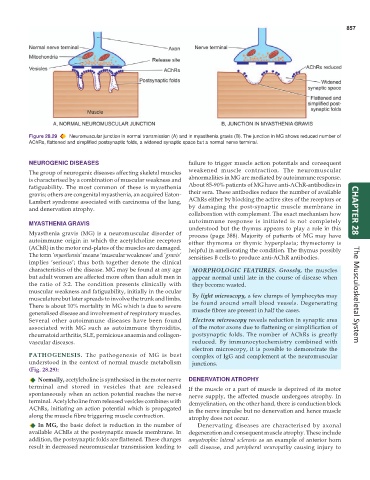
Figure 28.29 Neuromuscular junction in normal transmission (A) and in myasthenia gravis (B). The junction in MG shows reduced number of
AChRs, flattened and simplified postsynaptic folds, a widened synaptic space but a normal nerve terminal.
NEUROGENIC DISEASES failure to trigger muscle action potentials and consequent
The group of neurogenic diseases affecting skeletal muscles weakened muscle contraction. The neuromuscular
is characterised by a combination of muscular weakness and abnormalities in MG are mediated by autoimmune response.
fatiguability. The most common of these is myasthenia About 85-90% patients of MG have anti-AChR-antibodies in
gravis; others are congenital myasthenia, an acquired Eaton- their sera. These antibodies reduce the number of available
Lambert syndrome associated with carcinoma of the lung, AChRs either by blocking the active sites of the receptors or
and denervation atrophy. by damaging the post-synaptic muscle membrane in
collaboration with complement. The exact mechanism how CHAPTER 28
MYASTHENIA GRAVIS autoimmune response is initiated is not completely
understood but the thymus appears to play a role in this
Myasthenia gravis (MG) is a neuromuscular disorder of process (page 388). Majority of patients of MG may have
autoimmune origin in which the acetylcholine receptors either thymoma or thymic hyperplasia; thymectomy is
(AChR) in the motor end-plates of the muscles are damaged. helpful in ameliorating the condition. The thymus possibly
The term ‘myasthenia’ means ‘muscular weakness’ and ‘gravis’ sensitises B cells to produce anti-AChR antibodies.
implies ‘serious’; thus both together denote the clinical
characteristics of the disease. MG may be found at any age MORPHOLOGIC FEATURES. Grossly, the muscles
but adult women are affected more often than adult men in appear normal until late in the course of disease when
the ratio of 3:2. The condition presents clinically with they become wasted.
muscular weakness and fatiguability, initially in the ocular
musculature but later spreads to involve the trunk and limbs. By light microscopy, a few clumps of lymphocytes may The Musculoskeletal System
There is about 10% mortality in MG which is due to severe be found around small blood vessels. Degenerating
generalised disease and involvement of respiratory muscles. muscle fibres are present in half the cases.
Several other autoimmune diseases have been found Electron microscopy reveals reduction in synaptic area
associated with MG such as autoimmune thyroiditis, of the motor axons due to flattening or simplification of
rheumatoid arthritis, SLE, pernicious anaemia and collagen- postsynaptic folds. The number of AChRs is greatly
vascular diseases. reduced. By immunocytochemistry combined with
electron microscopy, it is possible to demonstrate the
PATHOGENESIS. The pathogenesis of MG is best complex of IgG and complement at the neuromuscular
understood in the context of normal muscle metabolism junctions.
(Fig. 28.29):
Normally, acetylcholine is synthesised in the motor nerve DENERVATION ATROPHY
terminal and stored in vesicles that are released If the muscle or a part of muscle is deprived of its motor
spontaneously when an action potential reaches the nerve nerve supply, the affected muscle undergoes atrophy. In
terminal. Acetylcholine from released vesicles combines with demyelination, on the other hand, there is conduction block
AChRs, initiating an action potential which is propagated in the nerve impulse but no denervation and hence muscle
along the muscle fibre triggering muscle contraction. atrophy does not occur.
In MG, the basic defect is reduction in the number of Denervating diseases are characterised by axonal
available AChRs at the postsynaptic muscle membrane. In degeneration and consequent muscle atrophy. These include
addition, the postsynaptic folds are flattened. These changes amyotrophic lateral sclerosis as an example of anterior horn
result in decreased neuromuscular transmission leading to cell disease, and peripheral neuropathy causing injury to