
Page 511 - First Aid for the USMLE Step 1 2020, Thirtieth edition [MedicalBooksVN.com]_Neat
P. 511
Musculoskeletal, skin, and connective tissue ` pathology Musculoskeletal, skin, and connective tissue ` pathology section iii 467
Gout
Findings Acute inflammatory monoarthritis caused by precipitation of monosodium urate crystals in
joints A . Risk factors: male sex, hypertension, obesity, diabetes, dyslipidemia, alcohol use.
Strongest risk factor is hyperuricemia, which can be caused by:
Underexcretion of uric acid (90% of patients)—largely idiopathic, potentiated by renal failure;
can be exacerbated by certain medications (eg, thiazide diuretics).
Overproduction of uric acid (10% of patients)—Lesch-Nyhan syndrome, PRPP excess, cell
turnover (eg, tumor lysis syndrome), von Gierke disease.
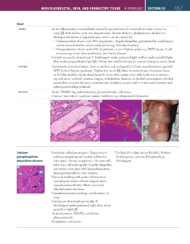
Crystals are needle shaped and ⊝ birefringent under polarized light (yellow under parallel light,
blue under perpendicular light B ). Serum uric acid levels may be normal during an acute attack.
symptoms Asymmetric joint distribution. Joint is swollen, red, and painful. Classic manifestation is painful
MTP joint of big toe (podagra). Tophus formation C (often on external ear, olecranon bursa,
or Achilles tendon). Acute attack tends to occur after a large meal with foods rich in purines
(eg, red meat, seafood), trauma, surgery, dehydration, diuresis, or alcohol consumption (alcohol
metabolites compete for same excretion sites in kidney as uric acid uric acid secretion and
subsequent buildup in blood).
tREatmEnt Acute: NSAIDs (eg, indomethacin), glucocorticoids, colchicine.
Chronic (preventive): xanthine oxidase inhibitors (eg, allopurinol, febuxostat).
A B C
Calcium Previously called pseudogout. Deposition of The blue P’s—blue (when Parallel), Positive
pyrophosphate calcium pyrophosphate crystals within the birefringence, calcium Pyrophosphate,
deposition disease joint space. Occurs in patients > 50 years old; Pseudogout
both sexes affected equally. Usually idiopathic,
A
sometimes associated with hemochromatosis,
hyperparathyroidism, joint trauma.
Pain and swelling with acute inflammation
(pseudogout) and/or chronic degeneration
(pseudo-osteoarthritis). Most commonly
affected joint is the knee.
Chondrocalcinosis (cartilage calcification) on
x-ray.
Crystals are rhomboid and weakly ⊕
birefringent under polarized light (blue when
parallel to light) A .
Acute treatment: NSAIDs, colchicine,
glucocorticoids.
Prophylaxis: colchicine.
FAS1_2019_11-Musculo.indd 467 11/7/19 5:23 PM