
Page 706 - First Aid for the USMLE Step 1 2020, Thirtieth edition [MedicalBooksVN.com]_Neat
P. 706
662 seCtioN iii RespiRatoRy ` RESPIRATORY—ANATOmY RespiRatoRy ` RESPIRATORY—ANATOmY
` RESPIRATORY—ANATOmY
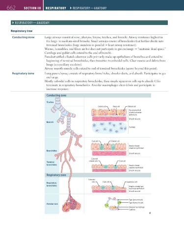
Respiratory tree
Conducting zone Large airways consist of nose, pharynx, larynx, trachea, and bronchi. Airway resistance highest in
the large- to medium-sized bronchi. Small airways consist of bronchioles that further divide into
terminal bronchioles (large numbers in parallel least airway resistance).
Warms, humidifies, and filters air but does not participate in gas exchange “anatomic dead space.”
Cartilage and goblet cells extend to the end of bronchi.
Pseudostratified ciliated columnar cells primarily make up epithelium of bronchus and extend to
beginning of terminal bronchioles, then transition to cuboidal cells. Clear mucus and debris from
lungs (mucociliary escalator).
Airway smooth muscle cells extend to end of terminal bronchioles (sparse beyond this point).
Respiratory zone Lung parenchyma; consists of respiratory bronchioles, alveolar ducts, and alveoli. Participates in gas
exchange.
Mostly cuboidal cells in respiratory bronchioles, then simple squamous cells up to alveoli. Cilia
terminate in respiratory bronchioles. Alveolar macrophages clear debris and participate in
immune response.
Conducting zone
Trachea
Goblet cell Basal cell Ciliated cell
Pseudostratified
ciliated columnar
epithelium
Smooth muscle
Bronchi
Cartilage
Club cell Ciliated cell
Simple ciliated
columnar epithelium
Bronchioles
Smooth muscle
Cuboidal
Terminal ciliated cells Club cell
bronchioles Simple ciliated
cuboidal epithelium
Smooth muscle
Respiratory zone
Cuboidal
Respiratory cells Club cell Squamous cell
bronchioles Simple cuboidal and
squamous epithelium
Smooth muscle
Type I pneumocyte
Type II pneumocyte
Alveolar sacs
Alveolar macrophage
Capillary
FAS1_2019_16-Respiratory.indd 662 11/8/19 7:34 AM