
Page 707 - First Aid for the USMLE Step 1 2020, Thirtieth edition [MedicalBooksVN.com]_Neat
P. 707
RespiRatoRy ` RESPIRATORY—ANATOmY RespiRatoRy ` RESPIRATORY—ANATOmY seCtioN iii 663
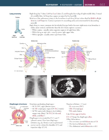
Lung anatomy Right lung has 3 lobes; Left has Less Lobes (2) and Lingula (homolog of right middle lobe). Instead
of a middle lobe, left lung has a space occupied by the heart A .
Trachea Upper lobe
Relation of the pulmonary artery to the bronchus at each lung hilum is described by RALS—Right
Anterior; Left Superior. Carina is posterior to ascending aorta and anteromedial to descending
Horizontal
aorta B . fissure Oblique fissure
Right lung is a more common site for inhaled foreign bodies because right main stem bronchus is
wider, more vertical, and shorter than the left. If you aspirate a peanut:
Middle lobe
Lingula
Carina While supine—usually enters superior segment of right lower lobe.
Lower
While lying on right side—usually enters right upper lobe. Lower
lobe
lobe
Inferior lobe
While upright—usually enters right lower lobe.
Right Left R L L R
bronchus bronchus Anterior view Posterior view
Anterior view Posterior view
Clavicle Midclavicular line
C7
1 T1
2
RUL 1 LUL 3 RUL
ICS1 Needle position 4
Horizontal fissure 2 for tension pneumothorax Oblique 5 T5 Horizontal
ICS2 LUL fissure fissure
3 6
RML RML
ICS3
4 LLL 7
Oblique fissure
Oblique fissure ICS4 8 T8 Oblique fissure
5
RLL ICS5 LLL 9 T9 RLL
6
10
7 11
Pleura Lingula 8 T12
9 12
10 L1
ICS = intercostal space
11
A B
RUL LUL
AAo
SVC PA
DAo
Carina
RML
RLL LLL
Diaphragm structures Structures perforating diaphragm: Number of letters = T level:
Inferior vena Central tendon At T8: IVC, right phrenic nerve T8: vena cava (IVC)
cava (T8) Esophagus At T10: esophagus, vagus (CN 10; 2 trunks) T10: (O)esophagus
(T10)
At T12: aorta (red), thoracic duct (white), T12: aortic hiatus
azygos vein (blue) (“At T-1-2 it’s the red, I ate (8) ten eggs at twelve.
white, and blue”)
Diaphragm is innervated by C3, 4, and 5 C3, 4, 5 keeps the diaphragm alive.
Rib (phrenic nerve). Pain from diaphragm Other bifurcations:
Aorta The common carotid bifourcates at C4.
(T12) irritation (eg, air, blood, or pus in peritoneal
Vertebra cavity) can be referred to shoulder (C5) and The trachea bifourcates at T4.
Inferior view The abdominal aorta bifourcates at L4.
trapezius ridge (C3, 4).
FAS1_2019_16-Respiratory.indd 663 11/8/19 7:34 AM