
Page 716 - First Aid for the USMLE Step 1 2020, Thirtieth edition [MedicalBooksVN.com]_Neat
P. 716
672 seCtioN iii RespiRatoRy ` RESPIRATORY—PAThOlOgY RespiRatoRy ` RESPIRATORY—PAThOlOgY
˙ ˙
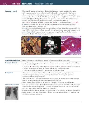
Pulmonary emboli V/Q mismatch, hypoxemia, respiratory alkalosis. Sudden-onset dyspnea, pleuritic chest pain,
tachypnea, tachycardia. Large emboli or saddle embolus A may cause sudden death due to
electromechanical dissociation (pulseless electrical activity). CT pulmonary angiography is
imaging test of choice for PE (look for filling defects) B . May have S1Q3T3 abnormality on ECG.
Lines of Zahn C are interdigitating areas of pink (platelets, fibrin) and red (RBCs) found only in
thrombi formed before death; help distinguish pre- and postmortem thrombi.
Types: Fat, Air, Thrombus, Bacteria, Amniotic fluid, Tumor. An embolus moves like a FAT BAT.
Fat emboli—associated with long bone fractures and liposuction; classic triad of hypoxemia,
neurologic abnormalities, petechial rash.
Air emboli—nitrogen bubbles precipitate in ascending divers (caisson disease/decompression sickness);
treat with hyperbaric O ; or, can be iatrogenic 2° to invasive procedures (eg, central line placement).
2
Amniotic fluid emboli—typically occurs during labor or postpartum, but can be due to uterine
trauma. Can lead to DIC. Rare, but high mortality.
A B C
Mediastinal pathology Normal mediastinum contains heart, thymus, lymph nodes, esophagus, and aorta.
Mediastinal masses Some pathologies (eg, lymphoma, lung cancer, abscess) can occur in any compartment, but there
are common associations:
Anterior—4T’s: Thyroid (substernal goiter), Thymic neoplasm, Teratoma, “Terrible” lymphoma.
Middle—esophageal carcinoma, metastases, hiatal hernia, bronchogenic cysts.
Posterior—neurogenic tumor (eg, neurofibroma), multiple myeloma.
Mediastinitis Inflammation of mediastinal tissues. Commonly due to postoperative complications of
cardiothoracic procedures (≤ 14 days), esophageal perforation, or contiguous spread of
odontogenic/retropharyngeal infection.
Chronic mediastinitis—also known as fibrosing mediastinitis; due to proliferation of connective
tissue in mediastinum. Histoplasma capsulatum is common cause.
Clinical features: fever, tachycardia, leukocytosis, chest pain, and sternal wound drainage.
Pneumomediastinum Presence of gas (usually air) in the mediastinum (black arrows show air around the aorta, red arrow
shows air dissecting into the neck A ). Can either be spontaneous (due to rupture of pulmonary
A
bleb) or 2° (eg, trauma, iatrogenic, Boerhaave syndrome).
Ruptured alveoli allow tracking of air into the mediastinum via peribronchial and perivascular sheaths.
Clinical features: chest pain, dyspnea, voice change, subcutaneous emphysema, ⊕ Hamman sign
(crepitus on cardiac auscultation).
FAS1_2019_16-Respiratory.indd 672 11/8/19 7:34 AM