Page 98 - First Aid for the USMLE Step 1 2020, Thirtieth edition [MedicalBooksVN.com]_Neat
P. 98

54          SECTION II    BIOCHEmISTRY  ``BIOCHEMISTRY—lABORATORY TECHNIqUES                                                                             BIOCHEmISTRY  ``BIOCHEMISTRY—lABORATORY TECHNIqUES


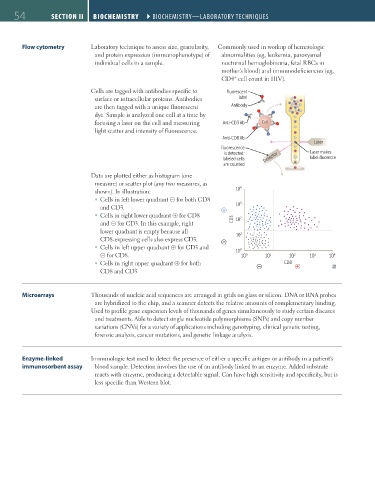
               Flow cytometry        Laboratory technique to assess size, granularity,   Commonly used in workup of hematologic
                                       and protein expression (immunophenotype) of   abnormalities (eg, leukemia, paroxysmal
                                       individual cells in a sample.            nocturnal hemoglobinuria, fetal RBCs in
                                                                                mother’s blood) and immunodeficiencies (eg,
                                                                                    +
                                                                                CD4  cell count in HIV).
                                     Cells are tagged with antibodies specific to   Fluorescent
                                       surface or intracellular proteins. Antibodies   label
                                       are then tagged with a unique fluorescent   Antibody
                                       dye. Sample is analyzed one cell at a time by
                                       focusing a laser on the cell and measuring   Anti-CD3 Ab  Cell
                                       light scatter and intensity of fluorescence.
                                                                                Anti-CD8 Ab
                                                                                                              Laser
                                                                                Fluorescence
                                                                                 is detected;                Laser makes
                                                                                labeled cells  Detector      label fluoresce
                                                                                 are counted
                                     Data are plotted either as histogram (one
                                       measure) or scatter plot (any two measures, as
                                       shown). In illustration:                      10 4
                                         ƒ Cells in left lower quadrant ⊝ for both CD8
                                        and CD3.                                     10 3
                                         ƒ Cells in right lower quadrant ⊕ for CD8    2
                                        and ⊝ for CD3. In this example, right       CD3  10
                                        lower quadrant is empty because all           1
                                        CD8-expressing cells also express CD3.       10
                                         ƒ Cells in left upper quadrant ⊕ for CD3 and   0
                                        ⊝ for CD8.                                   10 10 0  10 1    10 2   10 3  10 4
                                         ƒ Cells in right upper quadrant ⊕ for both                 CD8
                                        CD8 and CD3.


               Microarrays           Thousands of nucleic acid sequences are arranged in grids on glass or silicon. DNA or RNA probes
                                       are hybridized to the chip, and a scanner detects the relative amounts of complementary binding.
                                     Used to profile gene expression levels of thousands of genes simultaneously to study certain diseases
                                       and treatments. Able to detect single nucleotide polymorphisms (SNPs) and copy number
                                       variations (CNVs) for a variety of applications including genotyping, clinical genetic testing,
                                       forensic analysis, cancer mutations, and genetic linkage analysis.


               Enzyme-linked         Immunologic test used to detect the presence of either a specific antigen or antibody in a patient’s
               immunosorbent assay     blood sample. Detection involves the use of an antibody linked to an enzyme. Added substrate
                                       reacts with enzyme, producing a detectable signal. Can have high sensitivity and specificity, but is
                                       less specific than Western blot.


          FAS1_2019_01-Biochem.indd   54                                                                                11/7/19   3:16 PM
   93   94   95   96   97   98   99   100   101   102   103