
Page 123 - Template Tesis UTM v2.0
P. 123
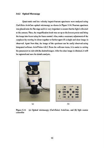
3.6.2  Optical Microscopy


                           Quasi-static and low velocity impact fracture specimens were analysed using

                   Carl-Zeiss AxioCam optical microscopy as shown in Figure 3.14. Fracture specimen

                   was placed onto the flat stage and it is very important to ensure that the light is directed
                   to the camera. Then, the magnification knob was set up to the lowest power and bring

                   the image into focus using the focus control. Also, make a necessary adjustment of the

                   eyepieces by moving its closer together or farther apart till a single and clear image is
                   observed. Apart from that, the image of the specimen can be easily observed using

                   integrated software AxioVision 4.8.2. From the software icons, it is easier to setting
                   the parameters to suit with the desired images. After the clear image is obtained, it will

                   be captured and save for details analysis.


                                     (a)                                      (b)


                   Figure 3.14    (a)  Optical  microscopy  (Carl-Zeiss)  AxioCam,  and  (b)  light  source
                   controller


                                                           93