
Page 287 - Hall et al (2015) Principles of Critical Care-McGraw-Hill
P. 287
CHAPTER 28: Interpretation of Hemodynamic Waveforms 191
II 0.5-20 Hz
1 mV
PAP [mm Hg] Ppa
60
54
48
42
36
30
24
18
12 Ppad
Whip artifact
6
0
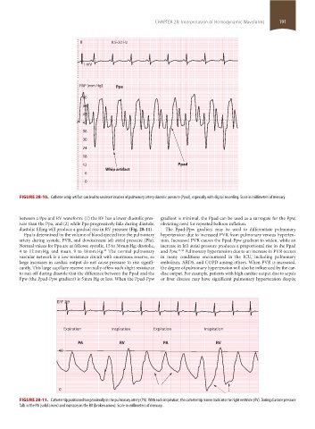
FIGURE 28-10. Catheter whip artifact can lead to underestimation of pulmonary artery diastolic pressure (Ppad), especially with digital recording. Scale in millimeters of mercury.
between a Ppa and RV waveform: (1) the RV has a lower diastolic pres- gradient is minimal, the Ppad can be used as a surrogate for the Ppw,
sure than the Ppa, and (2) while Ppa progressively falls during diastole, obviating need for repeated balloon inflation.
diastolic filling will produce a gradual rise in RV pressure (Fig. 28-11). The Ppad-Ppw gradient may be used to differentiate pulmonary
Ppa is determined by the volume of blood ejected into the pulmonary hypertension due to increased PVR from pulmonary venous hyperten-
artery during systole, PVR, and downstream left atrial pressure (Pla). sion. Increased PVR causes the Ppad-Ppw gradient to widen, while an
Normal values for Ppa are as follows: systolic, 15 to 30 mm Hg; diastolic, increase in left atrial pressure produces a proportional rise in the Ppad
4 to 12 mm Hg; and mean, 9 to 18 mm Hg. The normal pulmonary and Ppw. 33,34 Pulmonary hypertension due to an increase in PVR occurs
29
vascular network is a low-resistance circuit with enormous reserve, so in many conditions encountered in the ICU, including pulmonary
large increases in cardiac output do not cause pressure to rise signifi- embolism, ARDS, and COPD among others. When PVR is increased,
cantly. This large capillary reserve normally offers such slight resistance the degree of pulmonary hypertension will also be influenced by the car-
to run off during diastole that the difference between the Ppad and the diac output. For example, patients with high cardiac output due to sepsis
Ppw (the Ppad-Ppw gradient) is 5 mm Hg or less. When the Ppad-Ppw or liver disease may have significant pulmonary hypertension despite
ERF 2.9
Expiration Inspiration Expiration Inspiration
PA RV PA RV
40
0
FIGURE 28-11. Catheter tip positioned too proximally in the pulmonary artery (PA). With each inspiration, the catheter tip moves back into the right ventricle (RV). During diastole pressure
falls in the PA (solid arrow) and increases in the RV (broken arrow). Scale in millimeters of mercury.
section02.indd 191 1/13/2015 2:05:32 PM