
Page 368 - Hall et al (2015) Principles of Critical Care-McGraw-Hill
P. 368
238 PART 3: Cardiovascular Disorders
pulmonary vasoconstriction. In these circumstances, O therapy and Anaphylactic, Neurogenic, and Adrenal Shock: Other etiologies of shock
2
pulmonary vasodilator therapy combine to decrease pulmonary hyper- having unique clinical presentations that usually lead to early diagno-
tension and increase Q ˙ t in a small but significant proportion of patients sis are anaphylactic shock and neurogenic shock. Beyond identifying
(see Chap. 38). the etiology early through their association with triggering agents and
Right heart catheterization shows a unique hemodynamic profile: trauma, respectively, the physician should note that the pathophysiol-
a very high mean pulmonary artery pressure, pulmonary arterial DP ogy of each is a dilated venous bed with greatly increased unstressed
considerably greater than the Ppw and reduced Q ˙ t and SV. Not uncom- volume of the circulation leading to hypovolemic shock. Accordingly,
monly, arterial Ppw is normal or increased despite a small LVEDV on the mainstay of therapy for both conditions is adequate volume
echocardiographic examination, which also shows a right-to-left shift infusion; adjunctive therapy for anaphylaxis includes antihistamines,
of the interventricular septum; presumably, this causes stiffening of the steroids, and epinephrine to antagonize the mediators released in the
diastolic V-P curve of the left ventricle. A complication of pulmonary anaphylactic reaction (see Chap. 128), whereas a careful search for
vasodilator therapy is hypotension due to systemic arterial dilation sources of blood loss and hemorrhagic shock is part of the early resus-
unaccompanied by increased right heart output. Such effects aggravate citation of spinal shock in the traumatized patient (see Chap. 119).
the hypoperfusion state, perhaps by reducing coronary blood flow Not uncommonly, the presentation of patients with nonhemorrhagic
to the hypertrophied, dilated right ventricle. Some evidence suggests hypovolemic shock raises the concern of acute adrenal cortical insuffi-
that shock associated with pulmonary hypertension is ameliorated by ciency. When this possibility is not obviously excluded, it is appropriate
α-agonist therapy (eg, norepinephrine or phenylephrine), which acts as to draw a serum cortisol level, provide adequate circulating steroids with
a predominant systemic arteriolar constrictor to increase BP sufficiently dexamethasone, and conduct a corticotropin stimulation test to confirm
to maintain right ventricular perfusion. 59,60 or refute the diagnosis. Characteristically, hypotension and hypoperfu-
Right ventricular infarction causes low pulmonary artery pressures sion in such patients will not respond to adequate vascular volume
and normal LV filling pressures because the dilated, injured right ven- expansion until dexamethasone is administered (see Chap. 102).
tricle is unable to maintain adequate flow to the left heart. Elevated
61
neck veins and Pra tend to decrease with dobutamine infusion, perhaps Multiple Etiologies of Shock: With this differential diagnosis and man-
because the enhanced contractility of the left ventricle improves systolic agement evaluation in mind, the initial approach to patients with
function of the mechanically interdependent right ventricle. Volume hypoperfusion states should be completed in less time than it takes to
57
expansion often aggravates right ventricular dysfunction, and systemic read about it. The target is to distinguish among patients with septic
vasoconstriction may preserve right ventricular perfusion. 62 shock, cardiogenic shock, and hypovolemic shock and to initiate an
In the setting of severe AHRF, marked elevations of pulmonary appropriate therapeutic challenge—antibiotics, inotropic agents, or a
vascular resistance can be induced by hypoxic vasoconstriction. 63-65 volume challenge—within 30 minutes of presentation. By the response,
This hypoxic pulmonary vasoconstriction appears to be stimulated by the diagnosis is confirmed or challenged, with special regard to equivo-
mixed venous hypoxemia. In most patients with ARDS, this explains cal responses to therapy or to several other diagnostic categories of
66
increase. In a subset of these shock. Sorting out the primary etiology of the hypoperfusion state
the increase in Q ˙ s/Q ˙ t when CO and Pv O 2 often requires considerable additional data. This process is rendered
patients, RV dysfunction with dilatation of the RV and bowing of the
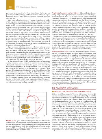
interventricular septum can be seen (Fig. 31-10). Treatment is similar more complex by concurrent etiologies contributing to the shock, for
63
as described above but additional therapies take advantage of the hetero- example, the patient with septic shock unable to increase Q ˙ t due to
geneity of Q ˙ s/Q ˙ t to deliver vasodilators directly to still ventilated alveoli intercurrent myocardial dysfunction, the patient with acute myocardial
and their accompanying vasculature. An example of this form of therapy infarction who is hypovolemic, or the patient with hemorrhagic shock
includes inhaled nitric oxide. Large multicenter trials, while demon- who becomes septic. Other combinations of these major categories
67
strating improvements in oxygenation early in the course of treatment, overlap with confounding effects of tamponade, positive-pressure ven-
did not demonstrate any mortality benefit. 68-70 tilation, pneumothorax, and pulmonary hypertension—all to challenge
ongoing diagnostic and management approaches.
THE PULMONARY CIRCULATION
■
120 PRESSURES, FLOW, AND RESISTANCE IN PULMONARY VESSELS
Q ˙ t from the left heart is equal to VR to the right heart, so the entire Q ˙ t
traverses the pulmonary circulation in pulsatile fashion (Fig. 31-11). The
Systolic 100 right ventricle ejects blood into the pulmonary artery, thereby increasing
its pressure (Ppa) to drive flow through a branching arteriolar system
80
Left ventricular pressure RV Distension into the lung parenchyma, where a network of very small alveolar septal
vessels or capillaries passes between the airspaces of the lung to effect
pulmonary gas exchange. These septal vessels converge into pulmonary
veins that empty into the left atrium, where the pressure (Pla) is often
Diastolic 20 RV LV A B regarded as the outflow pressure of the pulmonary circulation. When
this pressure gradient across the pulmonary circulation (Ppa − Pla) is
10
tance is calculated (mm Hg/L per minute) and sometimes converted to
Normal divided by the pulmonary blood flow (Q ˙ ), the pulmonary vascular resis-
5
50 100 150 metric units (dyn-s/cm ) by multiplying by 80. By this analysis, increas-
Left ventricular volume ing blood flow from one level to another is associated with decreasing
pressure across the pulmonary circulation (Ppa − Pla) along a unique
FIGURE 31-10. Systolic and diastolic volume-pressure (V-P) curves of the LV before pressure-flow relation given by the continuous line in Figure 31-11B.
(continuous curves) and during pulmonary hypertension (interrupted curve AB) in AHRF. This Resistance to Q ˙ may be increased by smooth muscle constriction within
LV diastolic dysfunction is due to RV distention and bowing of the interventricular septum (see the pulmonary arterioles and alveolar vessels by hypoxia, by compres-
inset cross-sectional diagram) so that the LV preload and SV are reduced. Pulmonary vasodila- sion of the alveolar septal vessels by elevated Pa, by obstruction of larger
tors such as NO have some therapeutic effect, but extracorporeal membrane oxygenation pulmonary vessels by thromboembolism, or by obliteration of many of
may provide better results. the parallel vascular channels as they traverse the lung so that the same
(ECMO) to increase Pv O 2
section03.indd 238 1/23/2015 2:06:44 PM