
Page 637 - Hall et al (2015) Principles of Critical Care-McGraw-Hill
P. 637
456 PART 4: Pulmonary Disorders
APPROACH TO DIAGNOSIS OF ALI AND ARDS Clinical Setting: The clinical setting in which the disorder develops can
■ CLINICAL PRESENTATION AND DIFFERENTIAL DIAGNOSIS often accompanied by systolic left ventricular or valvular dysfunction,
provide important diagnostic information. Cardiogenic edema is most
AHRF has many etiologies besides ALI and ARDS (Table 52-3). and the abnormal heart sounds and murmurs associated with each
However, the bedside appearance of patients with various forms of AHRF should be sought. Electrocardiographic (ECG) and serum enzyme evi-
dence of ischemia should be considered and suggest an obvious cause
is remarkably similar. Marked tachypnea and dyspnea are invariably
present. Physical examination reveals diffuse crackles in cases of cardio- for cardiogenic edema. Review of intravascular volume administration
often will supply information suggesting the explanation for pulmonary
genic pulmonary edema and focal findings of consolidation in cases of
lobar pneumonia. Cardiogenic pulmonary edema may be accompanied edema in patients with left ventricular or renal dysfunction.
ALI and ARDS commonly arise in a typical clinical context (see
by evidence of airflow obstruction, including wheezing and hypercap-
nia. The presence of crackles, a radiologic appearance of high-pressure Table 52-2). Sepsis, pneumonia, trauma, transfusion of blood prod-
215
ucts, and acid aspiration account for the majority of cases of ALI and
edema (see below), and hypoxemia refractory to oxygen therapy, all 3,4,13,38
suggest cardiogenic pulmonary edema as the primary process. Cough ARDS. Less common causes include pancreatitis, near-drowning,
leukoagglutination reactions, lung infections with viral agents or
and purulent sputum are hallmarks of infectious processes, while copious 38
clear or pink-colored airway secretions result from fulminant (“flash”) Pneumocystis jiroveci, fat embolism syndrome, and drug toxicities.
cardiogenic pulmonary edema. Chest Radiograph: The chest radiograph is a simple and widely available
Distressed patients with AHRF typically have initial room air arte- test used to assess patients with AHRF. Unfortunately, the accuracy of
in the 30 to 55 mm Hg range and pulse
rial blood gas results with Pa O 2 the routine radiograph in distinguishing hydrostatic from increased
oximetry less than 85% of arterial O saturation. If supplemental oxygen permeability edema is not high. 216,217 Criteria that have been suggested
2
by mask or cannula raises arterial saturation to above 95%, a large intra- to support a diagnosis of hydrostatic edema include increased heart size,
pulmonary shunt is unlikely. Other causes of respiratory distress should increased width of the vascular pedicle, vascular redistribution toward
then be considered, including airways disease, pulmonary embolus, or upper lobes, septal lines, and a centrifugal pattern of spread with a peri-
severe metabolic acidosis. Failure to achieve >95% saturation of arterial hilar bat’s-wing distribution of the edema. The lack of these findings and
blood with supplemental oxygen indicates the presence of a large right- patchy peripheral infiltrates that extend to the lateral lung margins sug-
to-left shunt. The specific process should be investigated via physical gest ARDS. However, all these signs overlap, and in the best of hands this
examination and chest radiograph. In the rare instances that the chest test is unlikely to yield better than a 60% to 80% accuracy of diagnosis
radiograph is entirely clear of alveolar infiltrates, one should consider when applied without other diagnostic tools. 216
that the blood gas data are erroneous, that there is an anatomic right-
to-left shunt at another site (eg, pulmonary arteriovenous malforma- Echocardiography: Echocardiography is a useful noninvasive diagnostic
tions or intracardiac shunt), or that there is continued perfusion of an tool to obtain information regarding cardiovascular function 218,219 and
unventilated lung due to recent complete or nearly complete occlusion of may provide useful diagnostic and/or therapeutic information in ALI
its main bronchus (but before the lung has collapsed due to absorption patients. Left ventricular dilation, regional or global wall motion
220
atelectasis) (see Table 52-3). abnormalities, and substantial mitral regurgitation on Doppler imaging
The differential diagnosis of ALI and ARDS (ie, AHRF with diffuse support a diagnosis of cardiogenic edema. A heart with echocardio-
pulmonary infiltrates consistent with pulmonary edema in the absence graphically normal dimensions and function (both systolic and dia-
of a cardiac etiology) includes a variety of disorders and etiologies. stolic) in a patient with pulmonary edema suggests pulmonary vascular
Identifying the etiologies of the diffuse infiltrates is important because leakage, although prior ventricular or valvular dysfunction with inter-
specific treatments exist for several of these conditions (eg, acute eosino- current resolution of the high pulmonary vascular pressures predispos-
philic pneumonia or diffuse alveolar hemorrhage). Table 52-4 lists the ing to cardiogenic edema must be kept in mind.
major clinical and diagnostic characteristics of these disorders. In patients with ALI, pulmonary vascular dysfunction, as measured
by transpulmonary gradient or pulmonary vascular resistance index
derived from pulmonary artery catheter data, is common and predictive
of mortality. However, isolated echocardiography-derived measures of
221
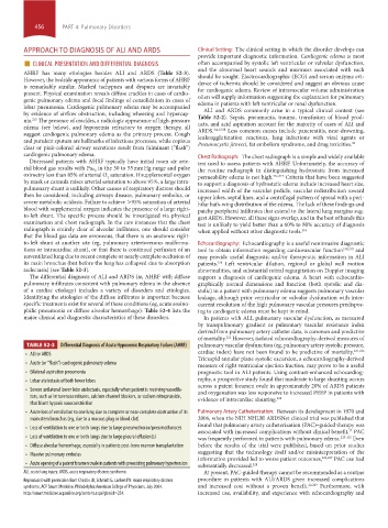
TABLE 52-3 Differential Diagnosis of Acute Hypoxemic Respiratory Failure (AHRF) pulmonary vascular dysfunction (eg, pulmonary artery systolic pressure,
• ALI or ARDS cardiac index) have not been found to be predictive of mortality, 221,222
Tricuspid annular plane systolic excursion, a echocardiography-derived
• Acute (or “flash”) cardiogenic pulmonary edema
measure of right ventricular ejection fraction, may prove to be a useful
• Bilateral aspiration pneumonia prognostic tool in ALI patients. Using contrast-enhanced echocardiog-
• Lobar atelectasis of both lower lobes raphy, a prospective study found that moderate to large shunting occurs
across a patent foramen ovale in approximately 20% of ARDS patients
• Severe unilateral lower lobe atelectasis, especially when patient is receiving vasodila- and oxygenation was less responsive to increased PEEP in patients with
tors, such as intravenous nitrates, calcium channel blockers, or sodium nitroprusside, evidence of intracardiac shunting. 220
that blunt hypoxic vasoconstriction
• Acute loss of ventilation to one lung due to complete or near-complete obstruction of its Pulmonary Artery Catheterization: Between its development in 1970 and
main stem bronchus (eg, due to a mucous plug or blood clot) 2006, when the NIH NHLBI ARDSNet clinical trial was published that
found that pulmonary artery catheterization (PAC)–guided therapy was
• Loss of ventilation to one or both lungs due to large pneumothorax/pneumothoraces
associated with increased complications without clinical benefit, PAC
22
• Loss of ventilation to one or both lungs due to large pleural effusion(s) was frequently performed in patients with pulmonary edema. 223-225 Even
• Diffuse alveolar hemorrhage, especially in patients post-bone marrow transplantation before the results of the trial were published, based on prior studies
• Massive pulmonary embolus suggesting that the technology itself and/or misinterpretation of the
information provided led to worse patient outcomes, 226,227 PAC use had
• Acute opening of a patent foramen ovale in patients with preexisting pulmonary hypertension substantially decreased. 228
ALI, acute lung injury; ARDS, acute respiratory distress syndrome. At present, PAC-guided therapy cannot be recommended as a routine
Reproduced with permission from Christie JD, Schmidt G, Lanken PN: Acute respiratory distress procedure in patients with ALI/ARDS given increased complications
syndrome, ACP Smart Medicine, Philadelphia:American College of Physicians, July 2004. and increased cost without a proven benefit. 22,229 Furthermore, with
http://smartmedicine.acponline.org/content.aspx?gbosid=234. increased use, availability, and experience with echocardiography and
section04.indd 456 1/23/2015 2:19:40 PM