
Page 638 - Hall et al (2015) Principles of Critical Care-McGraw-Hill
P. 638
CHAPTER 52: Acute Lung Injury and the Acute Respiratory Distress Syndrome 457
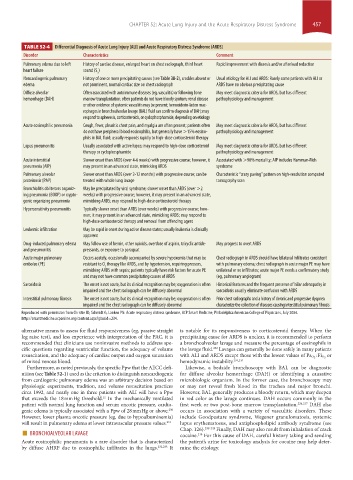
TABLE 52-4 Differential Diagnosis of Acute Lung Injury (ALI) and Acute Respiratory Distress Syndrome (ARDS)
Disorder Characteristics Comment
Pulmonary edema due to left History of cardiac disease, enlarged heart on chest radiograph, third heart Rapid improvement with diuresis and/or afterload reduction
heart failure sound (S )
3
Noncardiogenic pulmonary History of one or more precipitating causes (see Table 38-2), crackles absent or Usual etiology for ALI and ARDS: Rarely some patients with ALI or
edema not prominent, normal cardiac size on chest radiograph ARDS have no obvious precipitating cause
Diffuse alveolar Often associated with autoimmune diseases (eg, vasculitis) or following bone May meet diagnostic criteria for ARDS, but has different
hemorrhage (DAH) marrow transplantation; often patients do not have bloody sputum; renal disease pathophysiology and management
or other evidence of systemic vasculitis may be present; hemosiderin-laden mac-
rophages in bronchoalveolar lavage (BAL) fluid can confirm diagnosis of DAH; may
respond to apheresis, corticosteroids, or cyclophosphamide, depending on etiology
Acute eosinophilic pneumonia Cough, fever, pleuritic chest pain, and myalgia are often present; patients often May meet diagnostic criteria for ARDS, but has different
do not have peripheral blood eosinophilia, but generally have >15% eosino- pathophysiology and management
phils in BAL fluid; usually responds rapidly to high-dose corticosteroid therapy
Lupus pneumonitis Usually associated with active lupus; may respond to high-dose corticosteroid May meet diagnostic criteria for ARDS, but has different
therapy or cyclophosphamide pathophysiology and management
Acute interstitial Slower onset than ARDS (over 4-6 weeks) with progressive course; however, it Associated with >90% mortality; AIP includes Hamman-Rich
pneumonia (AIP) may present in an advanced state, mimicking ARDS syndrome
Pulmonary alveolar Slower onset than ARDS (over 2-12 months) with progressive course; can be Characteristic “crazy paving” pattern on high-resolution computed
proteinosis (PAP) treated with whole lung lavage tomography scan
Bronchiolitis obliterans organiz- May be precipitated by viral syndrome; slower onset than ARDS (over >2
ing pneumonia (BOOP) or crypto- weeks) with progressive course; however, it may present in an advanced state,
genic organizing pneumonia mimicking ARDS; may respond to high-dose corticosteroid therapy
Hypersensitivity pneumonitis Typically slower onset than ARDS (over weeks) with progressive course; how-
ever, it may present in an advanced state, mimicking ARDS; may respond to
high-dose corticosteroid therapy and removal from offending agent
Leukemic infiltration May be rapid in onset during active disease states; usually leukemia is clinically
apparent
Drug-induced pulmonary edema May follow use of heroin, other opioids, overdose of aspirin, tricyclic antide- May progress to overt ARDS
and pneumonitis pressants, or exposure to paraquat
Acute major pulmonary Occurs acutely, occasionally accompanied by severe hypoxemia that may be Chest radiograph in ARDS should have bilateral infiltrates consistent
embolus (PE) resistant to O therapy like ARDS, and by hypotension, requiring pressors, with pulmonary edema; chest radiograph in acute major PE may have
2
mimicking ARDS with sepsis; patients typically have risk factors for acute PE unilateral or no infiltrates; acute major PE needs a confirmatory study
and may not have common precipitating causes of ARDS (eg, pulmonary angiogram)
Sarcoidosis The onset is not acute, but its clinical recognition may be; oxygenation is often Historical features and the frequent presence of hilar adenopathy in
impaired and the chest radiograph can be diffusely abnormal sarcoidosis usually eliminate confusion with ARDS
Interstitial pulmonary fibrosis The onset is not acute, but its clinical recognition may be; oxygenation is often Prior chest radiographs and a history of chronic and progressive dyspnea
impaired and the chest radiograph can be diffusely abnormal characterize the collection of diseases causing interstitial pulmonary fibrosis
Reproduced with permission from Christie JD, Schmidt G, Lanken PN: Acute respiratory distress syndrome, ACP Smart Medicine, Philadelphia:American College of Physicians, July 2004.
http://smartmedicine.acponline.org/content.aspx?gbosid=234.
alternative means to assess for fluid responsiveness (eg, passive straight is notable for its responsiveness to corticosteroid therapy. When the
leg raise test), and less experience with interpretation of the PAC, it is precipitating cause for ARDS is unclear, it is recommended to perform
recommended that clinicians use noninvasive methods to address spe- a bronchoalveolar lavage and measure the percentage of eosinophils in
cific questions regarding ventricular function, the adequacy of volume the lavage fluid. Lavages can generally be done safely in many patients
232
resuscitation, and the adequacy of cardiac output and oxygen saturation with ALI and ARDS except those with the lowest values of Pa O 2 : Fi O 2 or
of mixed venous blood. hemodynamic instability. 234,235
Furthermore, as noted previously, the specific Ppw that the AECC defi- Likewise, a bedside bronchoscopy with BAL can be diagnostic
nition (see Table 52-1) used as the criterion to distinguish noncardiogenic for diffuse alveolar hemorrhage (DAH) or identifying a causative
from cardiogenic pulmonary edema was an arbitrary decision based on microbiologic organism. In the former case, the bronchoscopy may
physiologic experiments, tradition, and volume resuscitation practices or may not reveal fresh blood in the trachea and major bronchi.
circa 1992, and nearly one in three patients with ALI will have a Ppw However, BAL generally produces a bloody return, which may deepen
that exceeds the 18 mm Hg threshold. In the mechanically ventilated in red color as the lavage continues. DAH occurs commonly in the
22
patient with normal lung function and serum oncotic pressure, cardio- first week or two post-bone marrow transplantation. 236,237 DAH also
genic edema is typically associated with a Ppw of 28 mm Hg or above. occurs in association with a variety of vasculitic disorders. These
230
However, lower plasma oncotic pressure (eg, due to hypoalbuminemia) include Goodpasture syndrome, Wegener granulomatosis, systemic
will result in pulmonary edema at lower intravascular pressure values. 231 lupus erythematosus, and antiphospholipid antibody syndrome (see
■ BRONCHOALVEOLAR LAVAGE Chap. 126). 238-243 Finally, DAH may also result from inhalation of crack
cocaine. For this cause of DAH, careful history taking and sending
238
Acute eosinophilic pneumonia is a rare disorder that is characterized the patient’s urine for toxicology analysis for cocaine may help deter-
by diffuse AHRF due to eosinophilic infiltrates in the lungs. 232,233 It mine the etiology.
section04.indd 457 1/23/2015 2:19:40 PM