
Page 130 - The Netter Collection of Medical Illustrations - Integumentary System_ Volume 4 ( PDFDrive )
P. 130
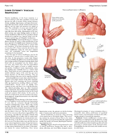
Plate 4-45                                                                                            Integumentary System

       LOWER EXTREMITY VASCULAR                                              VASCULAR INSUFFICIENCY IN DIABETES
       INSUFFICIENCY


       Vascular  insufficiency  of  the  lower  extremity  is  a   Dependent rubor,
       common finding in the older population. Factors that     absence of dorsalis
       increase  the  risk  of  vascular  disease  include  diabetes,   pedis pulsation
       obesity, smoking, hypertension, and hypercholesterol-
       emia. Both the venous and the arterial systems may be
       affected,  and  the  signs  and  symptoms  are  unique  to
       each. The combination of venous and arterial insuffi-
       ciency  is  commonly  seen  in  older  diabetic  patients,
       especially those who smoke. Abnormalities of the lym-
       phatic  system  may  cause  findings  similar  to  those  of
       venous insufficiency. Risk factors for lymphatic disease
       include prior surgeries (e.g., inguinal lymph node dis-
       section), radiotherapy, and idiopathic lymphedema.                                                Diabetic ulcer
         Clinical Findings: Venous insufficiency is a common
       disease that has no racial or ethnic predilection. It has
       been reported to be slightly more common in women.
       Venous  insufficiency  eventually  leads  to  venous  stasis
       and ulcerations. It has been estimated to be the cause
       of more than 50% of lower extremity ulcerations, with
       arterial  insufficiency  being  the  next  most  common
       cause,  and  neuropathic  causes  and  lymphedema
       accounting for the remainder.
         The  first  signs  of  venous  insufficiency  may  be  the
       development of varicose veins or smaller dilated retic-
       ular  veins.  As  time  progresses,  venous  stasis  changes
       are seen, including dry, pink to red, eczematous patches
       with varying amounts of peripheral pitting edema. Red                                 Valve
       blood  cells  are  extravasated  into  the  dermis  where,
       over  time,  they  break  down  and  form  hemosiderin
       deposits,  which  appear  as  brown  to  reddish  macules                          Healthy
       and  patches.  Continued  venous  hypertension,  stasis,                           vein
       and  swelling  may  eventually  lead  to  a  venous  stasis
       ulcer. These ulcers are most commonly present on the
       medial  malleolus  region  of  the  ankle  but  can  occur
       almost  anywhere  on  the  lower  extremity.  They  are   Extensive
       usually nontender, but some can be exquisitely painful.  gangrene
         Arterial insufficiency is most often caused by athero-
       sclerosis of the larger arteries of the lower extremity.
       Patients  often  have  coexisting  risk  factors  including
       older age, hypertension, smoking, diabetes, and hyper-
       cholesterolemia.  Arterial  ulcers  are  slightly  more
       common  in  men,  and  there  is  no  racial  predilection.
       The  clinical  presenting  signs  are  often  dependent
       rubor, claudication, and rest pain. Physical examination
       confirms the absence of peripheral pulses in the dorsal
       pedal  and  posterior  tibial  arteries.  At  this  point,  the
       patient is at high risk for arterial ulcerations and subse-
       quent gangrene. Surgical intervention is the only viable
       means of treatment.
         Pathogenesis: Venous drainage of the lower extrem-
       ity is accomplished via the superficial and deep systems                                                 Ulcer with lymphedema
       of  veins  that  are  connected  through  horizontally   Gangrene of toe                                 and stasis dermatitis
       arranged communicating vessels. These veins contain                                                      skin changes
       one-way  bicuspid  valves  that  prevent  backflow  and
       work with the action of muscle contraction to force the
       venous flow in a superior direction, eventually to empty
       into the inferior vena cava. The flow of venous blood   hypertension occurs, the patient is at risk for develop-  Histological evaluation of venous ulcerations shows a
       toward the vena cava is the primary responsibility of the   ment of venous stasis and venous ulcerations.  nonspecific  ulcer,  edema,  proliferation  of  superficial
       leg  muscles,  especially  the  calf  muscle.  Patients  with   Arterial insufficiency is caused by a slow narrowing   dermal vessels, and extravasated red blood cells with a
       sedentary lifestyles are at higher risk for venous insuf-  of the arteries due to cholesterol plaque. This narrow-  varying amount of hemosiderin deposition.
       ficiency. During ambulation, the venous pressure nor-  ing  restricts  the  amount  of  blood  flow  to  the  tissue.   Treatment:  Venous  insufficiency  is  treated  with  a
       mally decreases as the blood flow is increased toward   Once the flow is decreased to less than the requirement   combination of compression and leg elevation. Losing
       the vena cava. If an abnormality exists and this does not   needed for muscle and normal physiological function-  weight and increasing the activity level may also help.
       occur, venous hypertension ensues. Congenital absence   ing, symptoms arise.        Arterial insufficiency is best treated surgically with stent
       of the venous valves, incompetent valves, and a history   Histology: Biopsies should not be performed in cases   placement  or  arterial  bypass  of  the  narrowed  artery.
       of deep venous thrombosis are just three of the poten-  of  arterial  insufficiency,  because  they  lead  to  ulcer-  Pentoxifylline has also been used, with variable success,
       tial  reasons  for  venous  insufficiency.  Once  venous   ations,  infections,  and, most  likely, emergent surgery.   in early disease.

       116                                                                                   THE NETTER COLLECTION OF MEDICAL ILLUSTRATIONS