
Page 131 - The Netter Collection of Medical Illustrations - Integumentary System_ Volume 4 ( PDFDrive )
P. 131
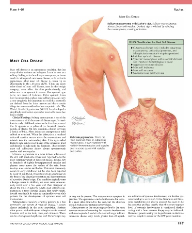
Plate 4-46                                                                                                           Rashes

                                                                                      MAST CELL DISEASE

                                                                              Solitary mastocytoma with Darier’s sign. Solitary mastocytomas
                                                                              almost always self-resolve. Darier’s sign is elicited by rubbing
                                                                              the mastocytoma, causing urtication.


                                                                                             WHO Classification for Mast Cell Disease

                                                                                                Cutaneous disease only (includes cutaneous
                                                                                                mastocytoma, urticaria pigmentosa, and
                                                                                                telangiectasia macularis eruptiva perstans)
                                                                                                Indolent systemic disease
                                                                                                Systemic mastocytosis with associated clonal
        MAST CELL DISEASE                                                                       non–mast cell hematological disease
                                                                                                Aggressive systemic disease
                                                                                                Mast cell leukemia
        Mast cell disease is an uncommon condition that has                                     Mast cell sarcoma
        many clinical variants and subtypes. It can be seen as a                                Noncutaneous mastocytoma
        solitary finding, as in the solitary mastocytoma, or it can
        result in widespread cutaneous disease, as in urticaria
        pigmentosa.  Most  mast  cell  disease  is  caused  by  an
        abnormality in the c-kit gene (KIT) . There are many
        other  forms  of  mast  cell  disease,  most  in  the  benign
        category;  some  affect  the  skin  predominantly,  and
        others are more systemic in nature. One systemic type
        is  the  rare  mast  cell  leukemia.  Other  systemic  forms
        have been reported, such as mast cell sarcoma, and carry
        a poor prognosis. It is important to recall that mast cells
        are  derived  from  the  bone  marrow  and  share  certain
        things in common with other hematopoietic cells. The
        World  Health  Organization  (WHO)  has  developed  a
        simplified classification system for mast cell disease (see
        box to right).
          Clinical Findings: Solitary mastocytoma is one of the
        most common of all the mast cell disease types. It mani-
        fests in early childhood, often in the first few years of
        life.  It  appears  as  a  yellowish  to  brownish  macule,
        papule, or plaque. On rare occasions, a lesion develops
        a vesicle or bulla. Most lesions are asymptomatic until
        rubbed or scratched. When this takes place, a localized
        urticarial reaction occurs above the mastocytoma and   Urticaria pigmentosa. This is the
        extends  into  the  surrounding  skin.  This  sign,  called   most common form of cutaneous
        Darier’s sign, can be used in any of the cutaneous mast   mastocytosis. It can manifest with
        cell diseases to help make the diagnosis. These solitary   reddish-brown macules and papules
        mast  cell  collections  almost  always  spontaneously   and in severe cases with vesicles
        resolve with no sequelae.                  and bullae.
          Urticaria pigmentosa is a more diffuse affliction of
        the skin with mast cells; it has been reported to be the
        most common variant of mast cell disease. From a few
        to  hundreds  of  slightly  hyperpigmented  macules  and
        plaques  occur  across  the  surface  of  the  skin.  Some
        develop into vesicles and bullae. This most commonly
        occurs  in  early  childhood  but  has  also  been  reported
        to occur in adulthood. Most children are diagnosed on
        the basis of the clinical presentation and demonstration
        of a positive Darier’s sign. The condition typically runs
        a benign course in children, and most cases spontane-
        ously  remit  over  a  few  years  and  then  disappear  at
        about  the  time  of  puberty.  Adult-onset  urticaria  pig-
        mentosa  is  a  more  chronic  disease  that  rarely  remits.
        Special care should be taken to continually screen adult
        patients  for  the  development  of  systemic  mast  cell   or may not be present. The most common symptom is   are indicative of systemic involvement, and further sys-
        involvement.                              pruritus. The appearance can be bothersome for some.   temic workup is warranted. Urine histamine and hista-
          Telangiectasia  macularis  eruptiva  perstans  is  a  less   It  is  most  often  limited  to  the  skin,  but  the  clinician   mine metabolites can also be assessed but seem to be
        commonly seen variant of mast cell disease. It occurs   should evaluate for systemic involvement.  less sensitive and less specific than the serum tryptase
        almost  exclusively  in  the  adult  population.  Patients   Measurement of the serum tryptase level is the most   level.  If  systemic  involvement  is  considered,  further
        often present with widespread telangiectases in unusual   accurate means of screening for systemic involvement   testing with a bone marrow biopsy may be indicated.
        locations such as the back, chest, and abdomen. There   with mastocytosis. Levels in the normal range indicate   Molecular genetic testing can be performed on the bone
        can be a background erythema, and Darier’s sign may   cutaneous  disease  only;  levels  greater  than  20 ng/mL   marrow sample to assess for the KIT gene mutation.


        THE NETTER COLLECTION OF MEDICAL ILLUSTRATIONS                                                                          117