
Page 127 - The Netter Collection of Medical Illustrations - Integumentary System_ Volume 4 ( PDFDrive )
P. 127
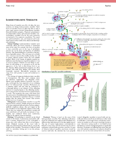
Plate 4-42                                                                                                           Rashes

                                                                                                  Circulating ANCA
                                                                     Cytokine receptor
                                                         Fc receptor               Circulating cytokine
                                                                         ANCA             Circulating cytokines migrate to cytokine receptors
                                                                         antigen          on neutrophil surface.

        LEUKOCYTOCLASTIC VASCULITIS
                                                                                    Cytokine primary results in expression of ANCA antigens on
                                                    Adhesion molecule               neutrophil cell surface.
        Many forms of vasculitis can affect the skin, the most                   Cytokine–cytokine receptor complex
        common  one  being  leukocytoclastic  vasculitis.  Other
        forms of vasculitis known to affect the skin as well as   Surface ANCA                  Circulating ANCA–ANCA antigen complex
        other  organ  systems  include  Churg-Strauss  vasculitis,   antigen, F(ab’) 2
        Henoch-Schönlein  purpura,  Wegener’s  granulomato-
        sis, polyarteritis nodosa, and urticarial vasculitis. Leu-  ANCA–ANCA antigen complex  Neutrophils activated by direct ANCA-F(ab’) 2  binding and by
        kocytoclastic  vasculitis  is  by  far  the  most  commonly   bound to Fc receptor  binding of ANCA–ANCA antigen complexes to Fc receptors
        encountered of the cutaneous vasculitides. The causes
        and  pathomechanisms  vary,  and  diagnosis  and  treat-                           Adhesion of neutrophils to vascular surface by interaction
        ment depend on the results of the clinical and histologi-                          of adhesion molecules with endothelial adhesion
        cal evaluations.                                                                   molecule receptors
          Clinical  Findings:  Leukocytoclastic  vasculitis  most                             Subendothelial migration of neutrophils
        commonly  affects  the  lower  extremity  or  dependent
        areas of the body. For example, this form of vasculitis                                     Apoptosis of endothelial cell
        is  most  commonly  seen  on  the  legs  of  ambulatory   Adhesion molecule–receptor complex
        patients  but  on  the  back  and  buttocks  of  bedridden   Endothelial cell
        patients. The clinical hallmark of vasculitis is the pres-
        ence of palpable purpura. The rash may start as small,
        pink, violaceous macules that rapidly develop into red
        or  purple  palpable  papules;  hence  the  term  palpable
        purpura.  Most  of  the  lesions  of  palpable  purpura  are   Process of neutrophil (or monocyte)
        uniform in size, but they can range from minute to 1 cm   activation by ANCA ultimately results
        or more in diameter. Patients are most likely to com-   in endothelial cell and neutrophil
        plain  of  mild  itching  or  no  symptoms  at  all,  and  the   apoptosis and necrosis with lytic
        appearance  of  the  rash  is  what  brings  them  to  see     disruption of vessel wall matrix.
        the  clinician.  Mild  constitutional  symptoms  are  often             Vessel wall      Neutrophil
                                                                                                 apoptosis and
        present,  with  mild  fever,  fatigue,  and  malaise  most                               necrosis
        commonly  reported.  Skin-specific  symptoms  can     Distribution of specific vasculitis syndromes
        range  from  mild  pruritus  to  pain  and  tenderness  to
        palpation.                                                                             Aorta
          The etiology of cutaneous leukocytoclastic vasculitis
        is  heterogeneous.  The  three  most  common  causes                   Large vessel
        are  infections,  medications,  and  idiopathic  causes.                 vasculitis  (giant cell  arteritis/  Takayasu  arteritis)
        Almost every possible infection (bacterial, viral, para-
        sitic, and fungal) has been reported to be an initiating   Medium-size  (polyarteritis
        factor  for  leukocytoclastic  vasculitis.  Medications  are     vessel  vasculitis  nodosa/  Kawasaki  disease)  Arteries
        a  common  culprit  and  can  easily  be  overlooked  if
        a  thorough  history  is  not  obtained.  If  the  offending
        infection is treated properly or the offending medica-
        tion is removed, the vasculitis resolves in approximately
        1 month. The symptoms also cease, often faster than                          Arterioles
        the rash resolves. Postinflammatory hyperpigmentation   Small vessel vasculitis (microscopic polyangiitis/Wegener’s granulomatosis)
        with  some  hemosiderin  deposition  often  is  a  residual                  Capillaries    Goodpasture’s   syndrome
        finding  after  the  lesions  have  cleared.  This  resolves                                                          granulomatosis/Churg-Strauss syndrome
        slowly over 6 to 12 months.                                                 Venules                                 Microscopic polyangiitis/Wegener’s
          Pathogenesis: Leukocytoclastic vasculitis is a type III                                                  Henoch-Schönlein purpura Cryoglobulinemia  ANCA small vessel vasculitis
        hypersensitivity reaction. Soluble antigens are believed                                           Isolated cutaneous leukocytoclastic angiitis
        to become complexed with antibodies. As these antigen-                    Veins
        antibody  complexes  enlarge,  they  get  trapped  in  the
        tiny vasculature of the dependent regions of the body.
        There, they can initiate the complement cascade and
        cause endothelial cell wall death, recruitment of neu-
        trophils,  and  continued  blood  vessel  destruction,
        leading to the typical cutaneous findings.
          Histology: The pathology is centered on the blood   Treatment:  Therapy  is  based  on  the  cause  of  the   treated.  Idiopathic  vasculitis  is  treated  with  oral  ste-
        venules in the dermis. A prominent neutrophilic infil-  leukocytoclastic vasculitis. New offending medications   roids, and often a search for an infection or other cause
        trate  is  present.  Degeneration  of  the  neutrophils  is   should be withdrawn and replaced with substitutes of a   is undertaken. A thorough history and physical exami-
        always seen, with nuclear dust; this is termed leukocy-  different class. Infections need to be thoroughly treated.   nation are needed, as well as some screening laboratory
        toclasis.  Fibrinoid  necrosis  of  the  vessel  walls  is   The  use  of  topical  high-potency  corticosteroids  is   tests.  Laboratory  testing  usually  is  not  helpful  unless
        easily seen. Extravasated red blood cells are seen in the   helpful in some cases, and oral steroids may be used in   the history or review of symptoms points in a particular
        vicinity of the vasculitis. Thrombosis of affected vessel   medication-induced leukocytoclastic vasculitis. In cases   direction. If patients are suffering from more than just
        walls  is  a  secondary  finding  and  is  not  the  primary   of  infection-induced  vasculitis,  prednisone  should  be   very mild systemic symptoms, an evaluation should be
        pathology.                                reserved  until  after  the  infection  has  been  properly   done to rule out the more serious forms of vasculitis.


        THE NETTER COLLECTION OF MEDICAL ILLUSTRATIONS                                                                          113