
Page 128 - The Netter Collection of Medical Illustrations - Integumentary System_ Volume 4 ( PDFDrive )
P. 128
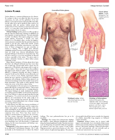
Plate 4-43                                                                                            Integumentary System

                                                               Generalized lichen planus
       LICHEN PLANUS                                                                                                   Classic lichen
                                                                                                                       planus. Purple,
                                                                                                                       polygonal, flat-
       Lichen planus is a common inflammatory skin disease.                                                            topped, pruritic
       It is unique in that it can affect the skin, the mucous                                                         papules
       membranes,  the  nails,  and  the  epithelium  of  the  hair
       follicles. Lichen planus most commonly affects the skin,
       but the other areas can be involved either solely or in
       conjunction  with  one  another.  Lichen  planus  that
       involves the skin has a tendency to spontaneously remit
       within 1 to 2 years after onset, whereas the oral version
       is almost always chronic in nature.
         Clinical Findings: Lichen planus can affect people at
       any age, but it is much more common in adulthood. It
       has no gender or racial predilection. The rash classically
       has been described as flat-topped, polygonal, pruritic,
       purple  papules.  Frequently,  a  whitish,  lacy  scale,
       referred  to  as  Wickham’s  striae,  overlies  the  papules.
       Lichen planus is unusual in that the pruritus causes the
       patient  to  rub  the  area,  rather  than  scratch.  Lichen
       planus  exhibits  the  Koebner  phenomenon,  and  often
       areas  of  linear  arrangement  are  seen  secondary  to
       trauma  or  rubbing.  This  is  helpful  when  clinically
       examining a patient, because scratch marks and excoria-
       tions  are  rarely  seen,  whereas  lichenification  from
       repeated rubbing of the lesions is frequently seen. The
       rash has a tendency to be more prominent on flexural
       surfaces,  especially  of  the  wrists.  The  glans  penis  is
       another  distinctive  location  in  which  lichen  planus
       commonly occurs.
         Many clinical variations of lichen planus have been
       described. An afflicted individual may have more than
       one  morphology.  Hypertrophic  lichen  planus  has  the
       appearance of thickened, scaly plaques with a rough or
       verrucal surface. There may be areas on the periphery
       that appear more classic in nature. This variant can be
       difficult  to  diagnosis  clinically,  and  often  a  biopsy  is
       required. It also can be difficult to treat effectively, and
       it  runs  a  chronic  course.  Rarely,  hypertrophic  lichen
       planus has been reported to transform into malignant
       squamous  cell  carcinoma.  Bullous  lichen  planus  is  an
       extremely uncommon variant that usually occurs on the
       lower extremities. The vesicle or bulla typically forms
       within the center of the lichen planus lesion.
         Lichen planopilaris is the term given to describe lichen
       planus affecting the terminal hair follicles. This is most
       common on the scalp and leads to a scarring alopecia.
       The  typical  findings  are  small,  erythematous  patches
       surrounding each hair follicle. As the disease progresses,
       loss of hair follicles is observed, signifying that scarring
       is taking place. The central crown is the area most often
       affected.  It  is  uncommon  for  the  entire  scalp  to  be   Oral lichen planus  Wickham’s striae. White  Histology of lichen planus.
       affected.  Once  scarring  has  occurred,  the  hair  loss  is             reticulated patches on the  Lichenoid lymphocytic
       permanent. Lichen planopilaris runs a chronic waxing                       buccal mucosa            infiltrate with “saw-toothing”
       and waning clinical course.                                                                         of the rete ridges, decreased
         Lichen planus may affect the mucous membranes of                                                  granular cell layer, and a
       the oral cavity, the genital region, and the conjunctiva.                                           Max Joseph space at the
       These  areas  appear  as  glistening  patches  with  lacy,                                          dermal-epidermal junction.
       white reticulations on the surface. Mucous membrane
       lichen planus has a higher tendency to ulcerate than the
       cutaneous  form  does.  There  have  been  reports  of
       malignant transformation to squamous cell carcinoma.
       For  this  reason,  long-term  follow-up  is  required.   findings.  The  exact  pathomechanism  has  yet  to  be   of eosinophils should lead one to consider the diagnosis
       Lichen planus may also affect the nail matrix and nail   described.                 of  a  lichen  planus–like  drug  eruption  or  lichenoid
       bed, leading to dystrophy and nail abnormalities. The   Histology:  The  lesions  show  characteristic  findings   contact dermatitis.
       most  frequently  seen  nail  abnormality  is  longitudinal   that  include  a  dense  lichenoid  lymphocytic  infiltrate   Treatment:  Isolated  lesions  can  be  treated  with
       ridging, but the most characteristic nail finding is pte-  along the dermal-epidermal border. Necrotic keratino-  topical corticosteroids. Up to two thirds of skin lesions
       rygium formation.                         cytes are frequently encountered within the hyperplas-  resolve spontaneously. Patients with widespread disease
         Pathogenesis: Lichen planus appears to be mediated   tic  epidermis  and  have  been  named  Civatte  bodies.   present a therapeutic challenge. Ultraviolet photother-
       by an abnormal T-cell immune response. The T cells   Hypergranulosis is a prominent feature as is the “saw-  apy,  oral  corticosteroids,  and  oral  retinoids  such  as
       act locally on the keratinocytes to induce the clinical   tooth” pattern of epidermal hyperplasia. The presence   acitretin and isotretinoin have been used.

       114                                                                                   THE NETTER COLLECTION OF MEDICAL ILLUSTRATIONS