
Page 135 - The Netter Collection of Medical Illustrations - Integumentary System_ Volume 4 ( PDFDrive )
P. 135
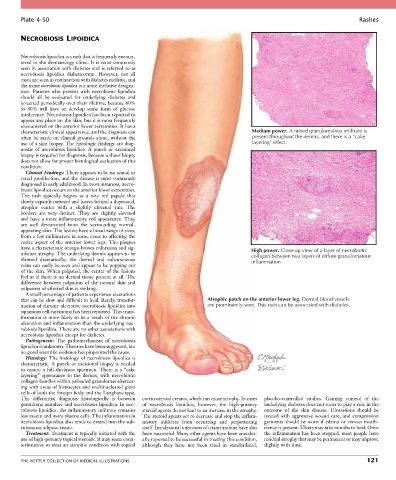
Plate 4-50                                                                                                           Rashes


        NECROBIOSIS LIPOIDICA


        Necrobiosis lipoidica is a rash that is frequently encoun-
        tered in the dermatology clinic. It is most commonly
        seen in association with diabetes and is referred to as
        necrobiosis  lipoidica  diabeticorum.  However,  not  all
        cases are seen in conjunction with diabetes mellitus, and
        the name necrobiosis lipoidica is a more inclusive designa-
        tion.  Patients  who  present  with  necrobiosis  lipoidica
        should  all  be  evaluated  for  underlying  diabetes  and
        screened periodically over their lifetime, because 60%
        to  80%  will  have  or  develop  some  form  of  glucose
        intolerance. Necrobiosis lipoidica has been reported to
        appear any place on the skin, but it is most frequently
        encountered on the anterior lower extremities. It has a
        characteristic clinical appearance, and the diagnosis can                       Medium power. A mixed granulomatous infiltrate is
        often be made on clinical grounds alone, without the                            present throughout the dermis, and there is a “cake
        use of a skin biopsy. The histologic findings are diag-                         layering” effect.
        nostic  of  necrobiosis  lipoidica.  A  punch  or  excisional
        biopsy is required for diagnosis, because a shave biopsy
        does not allow for proper histological evaluation of this
        condition.
          Clinical Findings: There appears to be no sexual or
        racial predilection, and the disease is most commonly
        diagnosed in early adulthood. In most instances, necro-
        biosis lipoidica occurs on the anterior lower extremities.
        The  rash  typically  begins  as  a  tiny  red  papule  that
        slowly expands outward and leaves behind a depressed,
        atrophic  center  with  a  slightly  elevated  rim.  The
        borders  are  very  distinct.  They  are  slightly  elevated
        and have a more inflammatory red appearance. They
        are  well  demarcated  from  the  surrounding  normal-
        appearing skin. The lesions have a broad range of sizes,
        from a few millimeters in some cases to affecting the
        entire  aspect  of  the  anterior  lower  legs.  The  plaques
        have a characteristic orange-brown coloration and sig-                          High power. Close-up view of a layer of necrobiotic
        nificant atrophy. The underlying dermis appears to be                           collagen between two layers of diffuse granulomatous
        thinned  dramatically;  the  dermal  and  subcutaneous                          inflammation
        veins can easily be seen and appear to be popping out
        of the skin. When palpated, the center of the lesions
        feel as if there is no dermal tissue present at all. The
        difference  between  palpation  of  the  normal  skin  and
        palpation of affected skin is striking.
          A small percentage of patients experience ulcerations
        that can be slow and difficult to heal. Rarely, transfor-        Atrophic patch on the anterior lower leg. Dermal blood vessels
        mation of chronic ulcerative necrobiosis lipoidica into          are prominently seen. This rash can be associated with diabetes.
        squamous cell carcinoma has been reported. This trans-
        formation is more likely to be a result of the chronic
        ulceration and inflammation than the underlying nec-
        robiosis lipoidica. There are no other associations with
        necrobiosis lipoidica except for diabetes.
          Pathogenesis:  The  pathomechanism  of  necrobiosis
        lipoidica is unknown. Theories have been suggested, but
        no good scientific evidence has pinpointed the cause.
          Histology: The histology of necrobiosis lipoidica is
        characteristic. A punch or excisional biopsy is needed
        to  ensure  a  full-thickness  specimen.  There  is  a  “cake
        layering”  appearance  to  the  dermis,  with  necrobiotic
        collagen bundles within palisaded granulomas alternat-
        ing with areas of histiocytes and multinucleated giant
        cells of both the foreign body and the Langhans type.
        The  differential  diagnosis  histologically  is  between   corticosteroid creams, which can cause atrophy. In cases   placebo-controlled  studies.  Gaining  control  of  the
        granuloma annulare and necrobiosis lipoidica. In nec-  of  necrobiosis  lipoidica,  however,  the  high-potency   underlying diabetes does not seem to play a role in the
        robiosis lipoidica, the inflammatory infiltrate contains   steroid agents do not lead to an increase in the atrophy.   outcome  of  the  skin  disease.  Ulcerations  should  be
        less mucin and more plasma cells. The inflammation in   The steroid agents act to decrease and stop the inflam-  treated  with  aggressive  wound  care,  and  compression
        necrobiosis lipoidica also tends to extend into the sub-  matory  infiltrate  from  occurring  and  perpetuating   garments should be worn if edema or venous insuffi-
        cutaneous adipose tissue.                 itself. Intralesional injections of triamcinolone have also   ciency is present. Ulcers may take months to heal. Once
          Treatment: Treatment is typically initiated with the   been successful. Many other agents have been anecdot-  the inflammation has been stopped, most people have
        use of high-potency topical steroids. It may seem coun-  ally reported to be successful in treating this condition,   residual atrophy that may be permanent or may improve
        terintuitive to treat an atrophic condition with topical   although  they  have  not  been  tried  in  standardized,   slightly with time.


        THE NETTER COLLECTION OF MEDICAL ILLUSTRATIONS                                                                          121