
Page 133 - The Netter Collection of Medical Illustrations - Integumentary System_ Volume 4 ( PDFDrive )
P. 133
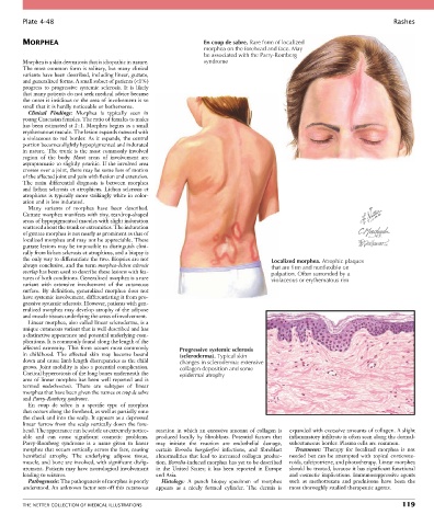
Plate 4-48                                                                                                           Rashes


        MORPHEA                                                  En coup de sabre. Rare form of localized
                                                                 morphea on the forehead and face. May
                                                                 be associated with the Parry-Romberg
        Morphea is a skin dermatosis that is idiopathic in nature.   syndrome
        The most common form is solitary, but many clinical
        variants have been described, including linear, guttate,
        and generalized forms. A small subset of patients (<1%)
        progress  to  progressive  systemic  sclerosis.  It  is  likely
        that many patients do not seek medical advice because
        the onset is insidious or the area of involvement is so
        small that it is hardly noticeable or bothersome.
          Clinical  Findings:  Morphea  is  typically  seen  in
        young Caucasian females. The ratio of females to males
        has been estimated at 2 : 1. Morphea begins as a small
        erythematous macule. The lesion expands outward with
        a violaceous to red border. As it expands, the central
        portion becomes slightly hypopigmented and indurated
        in nature. The trunk is the most commonly involved
        region  of  the  body.  Most  areas  of  involvement  are
        asymptomatic to slightly pruritic. If the involved area
        crosses over a joint, there may be some loss of motion
        of the affected joint and pain with flexion and extension.
        The  main  differential  diagnosis  is  between  morphea
        and  lichen  sclerosis  et  atrophicus.  Lichen  sclerosis  et
        atrophicus is typically more strikingly white in color-
        ation and is less indurated.
          Many  variants  of  morphea  have  been  described.
        Guttate morphea manifests with tiny, teardrop-shaped
        areas of hypopigmented macules with slight induration
        scattered about the trunk or extremities. The induration
        of guttate morphea is not nearly as prominent as that of
        localized morphea and may not be appreciable. These
        guttate lesions may be impossible to distinguish clini-
        cally from lichen sclerosis et atrophicus, and a biopsy is
        the only way to differentiate the two. Biopsies are not                       Localized morphea. Atrophic plaques
        always conclusive, and the term morphea–lichen sclerosis                      that are firm and nonflexible on
        overlap has been used to describe these lesions with fea-                     palpation. Often surronded by a
        tures of both conditions. Generalized morphea is a rare                       violaceous or erythematous rim
        variant  with  extensive  involvement  of  the  cutaneous
        surface.  By  definition,  generalized  morphea  does  not
        have systemic involvement, differentiating it from pro-
        gressive systemic sclerosis. However, patients with gen-
        eralized morphea may develop atrophy of the adipose
        and muscle tissues underlying the areas of involvement.
          Linear morphea, also called linear scleroderma, is a
        unique cutaneous variant that is well described and has
        a distinctive appearance and potential underlying com-
        plications. It is commonly found along the length of the
        affected extremity. This form occurs most commonly   Progressive systemic sclerosis
        in  childhood.  The  affected  skin  may  become  bound   (scleroderma). Typical skin
        down and cause limb length discrepancies as the child   changes in scleroderma: extensive
        grows. Joint mobility is also a potential complication.   collagen deposition and some
        Cortical hyperostosis of the long bones underneath the   epidermal atrophy
        area of linear morphea has been well reported and is
        termed  melorheostosis.  There  are  subtypes  of  linear
        morphea that have been given the names en coup de sabre
        and Parry-Romberg syndrome.
          En  coup  de  sabre  is  a  specific  type  of  morphea
        that occurs along the forehead, as well as partially onto
        the cheek and into the scalp. It appears as a depressed
        linear furrow from the scalp vertically down the fore-
        head. The appearance can be subtle or extremely notice-  reaction  in  which  an  excessive  amount  of  collagen  is   expanded with excessive amounts of collagen. A slight
        able  and  can  cause  significant  cosmetic  problems.   produced  locally  by  fibroblasts.  Potential  factors  that   inflammatory infiltrate is often seen along the dermal-
        Parry-Romberg  syndrome  is  a  name  given  to  linear   may  initiate  the  reaction  are  endothelial  damage,   subcutaneous border. Plasma cells are common.
        morphea that occurs vertically across the face, causing   certain  Borrelia  burgdorferi  infections,  and  fibroblast   Treatment:  Therapy  for  localized  morphea  is  not
        hemifacial  atrophy.  The  underlying  adipose  tissue,   abnormalities  that  lead  to  increased  collagen  produc-  needed  but  can  be  attempted  with  topical  corticoste-
        muscle, and bone are involved, with significant disfig-  tion. Borrelia-induced morphea has yet to be described   roids, calcipotriene, and phototherapy. Linear morphea
        urement. Patients may have neurological involvement   in  the  United  States;  it  has  been  reported  in  Europe   should be treated, because it has significant functional
        leading to seizures.                      and Asia.                                 and cosmetic implications. Immunosuppressive agents
          Pathogenesis: The pathogenesis of morphea is poorly   Histology:  A  punch  biopsy  specimen  of  morphea   such  as  methotrexate  and  prednisone  have  been  the
        understood. An unknown factor sets off this cutaneous   appears  as  a  nicely  formed  cylinder.  The  dermis  is   most thoroughly studied therapeutic agents.


        THE NETTER COLLECTION OF MEDICAL ILLUSTRATIONS                                                                          119