
Page 170 - The Netter Collection of Medical Illustrations - Integumentary System_ Volume 4 ( PDFDrive )
P. 170
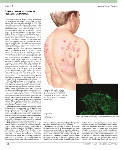
Plate 5-7                                                                                             Integumentary System

       LINEAR IMMUNOGLOBULIN A
       BULLOUS DERMATOSIS


       Linear immunoglobulin A (IgA) bullous dermatosis is
       an  infrequently  encountered  autoimmune  blistering
       disease  that  was  originally  described  in  1979.  This
       disease has a characteristic immunofluorescence stain-
       ing pattern that is used to differentiate it from other
       blistering diseases such as dermatitis herpetiformis. As
       the  name  implies,  linear  IgA  is  deposited  along  the
       length  of  the  dermal-epidermal  junction.  Chronic
       bullous disease of childhood is considered by most to
       be the same disease, although there are a few clinical
       differences in age at onset and associations that can be
       used to justify separating them into two distinct, albeit
       very similar, entities. Most cases of chronic bullous der-
       matosis of childhood are idiopathic, whereas most cases
       of linear IgA bullous dermatosis are drug induced and
       occur in an older population.
         Clinical Findings: Linear IgA bullous dermatosis is
       rare and is estimated to occur in 1 of every 2,000,000
       people. There is no race or sex predilection. It occurs
       most frequently in the adult population. The blistering
       disease has an insidious onset with small vesicles that
       may  mimic  dermatitis  herpetiformis.  The  blisters  are
       pruritic and do not have the same burning sensation as
       occurs in dermatitis herpetiformis, nor is there any rela-
       tionship  to  dietary  intake.  The  bullae  in  linear  IgA
       bullous dermatosis are characteristically arranged in a
       “string of sausages” configuration. Each bulla is elon-
       gated and tapers to an end, with a small area of inter-
       vening  normal-appearing  skin  before  the  tapering
       beginning of a new bulla. This string can be linear or
       annular in orientation. The blisters are tense and even-
       tually rupture and heal with minimal scarring. Mucous
       membrane  involvement  is  frequently  seen  and  can
       resemble that of mucous membrane pemphigoid.
         Chronic  bullous  disease  of  childhood  manifests  in
       early  childhood  (4-5  years  of  age).  The  blistering  is
       similar to that of linear Ig bullous dermatitis, and the
       histological findings are identical. Blistering in chronic
       bullous disease of childhood is more often localized to
       the abdomen and lower extremities but may occur any-  Characteristic bullae of linear
       where  on  the  skin;  it  also  commonly  affects  mucous   IgA disease, or chronic bullous
       membranes.  Chronic  bullous  disease  of  childhood  is   disease of childhood. They are
       most often idiopathic, whereas linear IgA bullous der-  configured in an annular manner
       matosis can also be seen in association with underlying   with small areas of intervening
       medications, malignancies, or other autoimmune con-  normal skin.
       ditions.  Many  medications  have  been  implicated  in
       causing linear IgA bullous dermatosis, with vancomycin
       being the most common by far.
         Histology: The immunofluorescence staining pattern
       is characteristic and shows linear IgA all along the base-
       ment membrane zone. This is highly specific and sensi-
       tive for the diagnosis of linear IgA bullous dermatosis
       and chronic bullous disease of childhood. Routine hema-
       toxylin and eosin staining shows a subepidermal blister
       with an underlying neutrophilic infiltrate. This can be                       Linear deposition of IgA along the basement membrane zone
       impossible to distinguish from dermatitis herpetiformis
       or bullous lupus, so immunostaining is required.
         Pathogenesis: The exact target antigen in linear IgA   various mechanisms, ultimately leading to disruption of   are  best  treated  by  recognizing  the  common  culprits
       bullous dermatosis is unknown. It is speculated that the   the dermal-epidermal junction and blistering.  and removing them immediately. Over a period of a few
       IgA  antibodies  are  directed  against  a  small  region  of   Treatment:  The  first  line  of  therapy  is  dapsone.   weeks, most patients who have discontinued the offend-
       bullous pemphigoid antigen 180 (BP180). Other pos-  Patients respond quickly to this medication. Low doses   ing medication return to a normal state. If the disease
       sible  antigens  exist  and  have  been  localized  to  the   of  dapsone  are  usually  all  that  is  needed.  Alternative   is found to be associated with an underlying malignancy
       lamina lucida and lamina densa regions of the basement   substitutes for dapsone include sulfapyridine and col-  or other autoimmune condition, therapy with dapsone
       membrane.  The  reason  for  formation  of  these  anti-  chicine.  Oral  prednisone  can  be  helpful  initially,  but   is  warranted.  Treatment  of  the  underlying  condition
       bodies  and  how  certain  medications  induce  them  are   because of the long-term side effects, patients should   should  also  be  undertaken.  If  the  malignancy  or  the
       unknown.  Once  present,  the  antibodies  target  the     be transitioned to one of the other medications men-  associated disease is put into remission, there is a good
       basement membrane zone and cause inflammation by   tioned. Drug-induced variants of this blistering disease   possibility that the blistering disease will remit as well.

       156                                                                                   THE NETTER COLLECTION OF MEDICAL ILLUSTRATIONS