
Page 169 - The Netter Collection of Medical Illustrations - Integumentary System_ Volume 4 ( PDFDrive )
P. 169
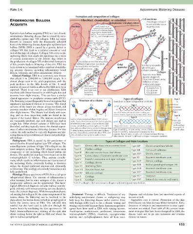
Plate 5-6                                                                                   Autoimmune Blistering Diseases

                                                       Formation and composition of collagen
        EPIDERMOLYSIS BULLOSA                              Fibroblast, chondroblast, or osteoblast    Cell membrane
                                                                               Endoplasmic reticulum cistern  Procollagen released
        ACQUISITA                                                                            Ribosome    to extracellular space
                                                    Nucleus          Hydroxylation of                    by pinocytosis
                                                                    certain prolyl and lysyl
                                                                   amino acid residues begins
        Epidermolysis bullosa acquisita (EBA) is a rare chronic     as pre-pro-  chains enter
                                                                 cistern. This requires vitamin C,
        autoimmune blistering disease that is caused by auto-      Fe , O , and  -ketoglutarate.
                                                                   2
                                                                     2
        antibodies  against  type  VII  collagen.  EBA  has  many
        features  in  common  with  the  dominantly  inherited   HO  HO OH  OH
        form of the blistering disease, dystrophic epidermolysis   HO  OH  OH  OH
        bullosa  (DEB).  DEB  is  caused  by  a  genetic  defect  in   OH  Gal  OH
        collagen  VII  that  leads  to  a  reduced  amount  or  total   Gal
        lack of this type of collagen. Collagen VII serves as the   OH  Gal  OH Gal Glc
        anchoring fibrils that attach the epidermis via a series   HO Gal
        of  protein  connections  to  the  dermis.  Any  defect  in     OH  Gal Glc  Gal
        the production of collagen VII or abnormal destruction      Glycosylation involves enzymatic
        of this protein leads to blistering of the skin. EBA has      addition of galactose to certain   Golgi   -s-s-  Terminal propeptides
                                                                                              -s-s- -s-s-
        been shown to be associated with a number of underly-    hydroxylysine residues by   apparatus  -s-s- -s-s-  split off by pro-
        ing  systemic  diseases,  including  inflammatory  bowel   galactosyltransferase.  -s-s- -s-s- -s-s-  -s-s-  collagen peptidase
        disease, leukemia, and other autoimmune diseases.            -s-s- -s-s-                                 Collagen
          Clinical Findings: EBA is an extremely rare disease   Gal  OH  -s-s-                        Assembly into fibrils
        that  affects  1  in  2,000,000  to  3,000,000  people.  It  is   OH  GalGlc      Disulfide   (quarter staggered).
                                                                                                      Cross links formed under
        almost  always  seen  in  the  adult  population,  with  the   GalGlc  OH  -s-s-    bonds     influence of lysyl
        peak  incidence  in  the  fifth  decade  of  life.  A  small   HO  Gal  -s-s-                 oxidase and copper
        number of cases of children affected by EBA have been   -s-s-  OH Three pro-   -s-s-             Gal galactose
                                                             chains assemble
        reported.  There  is  no  race  or  sex  predilection.  EBA   -s-s-  into triple helix,          Glc glucose
        manifests with blister formation or with fragile skin and   -s-s-  bonded by                     Gly glycine
        erosions  from  slight  trauma.  This  can  have  a  similar   OH groups.
        clinical appearance to porphyria cutanea tarda (PCT).                            Types of collagen                   Type I
        The blistering is most frequently located in regions that   Gly  Structure of   chains  Gly  (based on a chain composition of fibrils)  in
        experience mechanical friction or trauma. The dorsal     Gly                          Type I                        Bone
        surfaces of the hands are almost always involved, and                      1(I)                                     Tendon
        patients complain of skin fragility and blister formation                   2                                       Ligament
        after slight trauma. The blisters heal slowly with scar-                  Two  1(I) chains and one  2 chain   ( 1[I])   2;  Skin
                                                                                                         2
        ring,  and  on  close  inspection,  milia  are  found  in  the   X  Y  X  Y  in bone, tendon, ligament, fascia, skin, artery, uterus  Type II
        region of the healed blister. The mucous membranes   Each   chain comprises about 1,000 amino   Type II               in
        are  frequently  involved,  and  oral  disease  can  lead  to   acids. Every third amino acid in chain is glycine,   1(II)  Articular
        weight loss. Other clinical variants of EBA have been   smallest of amino acids. Glycine has no side                cartilage
        described, and they typically mimic the clinical appear-  chains, which thus permits tight coil. X and Y   Three  1(II) chains   ( 1[II]) 3 ; in articular cartilage  and cartilagi-
                                                                                                                            nous part of
        ance of other autoimmune blistering diseases. For this   here indicate other amino acids (X often proline;   Type III ( 1[III]) ; in skin, artery, uterus, GI tract.  growth plate
                                                   Y often hydroxyproline). Proline and hydroxy-
                                                                                           3
        reason, the only method to correctly diagnosis any blis-  proline, respectively, constitute about 10% and   Type IV ( 1[IV]) 3; in basement membranes, lens
        tering disease is by correlation of clinical and pathologi-  25% of total amino acids in each   chain.  capsule. Type V ( B) 3 or ( B)   A; in basement membranes,
                                                                                                   2
                                                                                 other tissues. At least 12 different collagen molecules identified.
        cal findings.
          Pathogenesis:  EBA  is  caused  by  the  production  of           Types of Collagen and Main Locations
        autoantibodies directed against type VII collagen. The
        noncollagenous portions of type VII collagen are the   Type I   Dermis, other tissue (most common form)  Type XII*  Dermis around hair follicles
        most antigenic sections. Type VII collagen is the main   Type II  Hyaline cartilage   Type XIII  Cell-to-cell adhesion
        component  of  the  anchoring  fibrils  found  within  the   Type III  Skin and vascular tissue, fetal dermis
        dermis. The antibodies that have been found are in the   Type IV     Lamina densa of basement membrane zone  Type XIV*  Dermis, cornea
        immunoglobulin  G  subclass.  They  activate  comple-                                 Type XV   Basement membrane zone
        ment, which results in inflammation and destruction of   Type V  Found in association with type I collagen  Type XVI*  Dermis, cartilage
        the  anchoring  fibrils,  eventually  leading  to  fractures   Type VI  Cartilage, dermis
        within the dermal-epidermal junction and, ultimately,   Type VII  Anchoring fibrils   Type XVII  Bullous pemphigoid antigen 180
        to blistering. The etiology of antibody formation is not   Type VIII  Vascular tissue, eye  Type XVIII  Basement membrane zone
        fully understood.                             Type IX*  Articular cartilage           Type XIX*  Basement membrane zone
          Histology: Biopsy specimens of EBA show a cell-poor
        subepidermal  blister.  The  amount  of  inflammation  is   Type X  Cartilage         Type XX *  Unknown
        often  minimal,  but  in  some  subtypes  of  the  disease  a   Type XI  Cartilage    Type XXI*  Extracellular vascular wall matrix
        lymphocytic  infiltrate  can  be  appreciated.  The  histo-  *FACIT collagen, fibril-associated collagens with interrupted triple helices.
        logical differential diagnosis includes bullous pemphi-
        goid, and only with immunostaining can one decisively
        make the correct diagnosis. With immunohistochemi-
        cal staining for collagen IV, the main component of the   Treatment:  Therapy  is  difficult.  Treatment  of  any   Dapsone and colchicine have had anecdotal reports of
        lamina  densa,  the  blistering  can  be  localized  to  the   underlying  autoimmune  disease  or  malignancy  may   success as well.
        plane above the lamina densa in bullous pemphigoid or   help  keep  the  blistering  disease  under  control.  Even   Supportive  care  is  critical.  Protection  of  the  skin
        below  the  lamina  densa  in  EBA.  The  salt-split  skin   with therapy, EBA tends to run a chronic waxing and   from trauma can help decrease blister formation. Early
        method  has  also  been  used  to  split  skin  through  the   waning course with frequent flares. Immunosuppressive   detection of infection and intervention to treat super-
        lamina lucida by incubating the skin specimen in 1M   agents  have  been  used  in  EBA  with  varying  success.   infection are critical. Even with all the current treat-
        NaCl.  Immunofluorescence  staining  of  the  split  skin   Azathioprine,  methotrexate,  prednisone,  intravenous   ment strategies that have been attempted for EBA, the
        shows staining below the split in EBA and above the   immunoglobulin  (IVIG),  rituximab,  mycophenolate   disease  tends  not  to  go  into  remission  and  remains
        split in bullous pemphigoid.              mofetil,  and  cyclophosphamide  have  all  been  used.   chronic in nature.


        THE NETTER COLLECTION OF MEDICAL ILLUSTRATIONS                                                                          155