
Page 168 - The Netter Collection of Medical Illustrations - Integumentary System_ Volume 4 ( PDFDrive )
P. 168
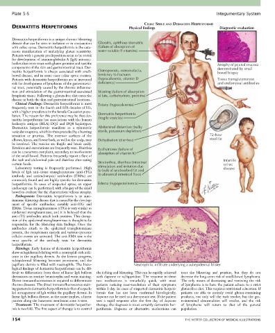
Plate 5-5                                                                                             Integumentary System

                                                                         CELIAC SPRUE AND DERMATITIS HERPETIFORMIS
       DERMATITIS HERPETIFORMIS                                  Physical findings                            Diagnostic evaluation


       Dermatitis herpetiformis is a unique chronic blistering
       disease that can be seen in isolation or in conjunction   Glossitis, aphthous stomatitis
       with celiac sprue. Dermatitis herpetiformis is the cuta-  (failure of absorption of
       neous  manifestation  of  underlying  gluten  sensitivity.   water-soluble B vitamins)
       Patients with a genetic predisposition seem to be at risk
       for development of immunoglobulin A (IgA) autoanti-
       bodies that cross-react with gluten proteins and specific                                             Atrophy of jejunal mucosa
       components of the skin and gastrointestinal tract. Der-                                               demonstrated by small
       matitis  herpetiformis  is  always  associated  with  small-  Osteoporosis, osteomalacia,             bowel biopsy
       bowel disease, and in some cases celiac sprue coexists.   tendency to fractures
       Patients with dermatitis herpetiformis are at increased   (hypocalcemia, vitamin D                    Tissue transglutaminase
       risk for development of lymphoma of the gastrointesti-  deficiency)                                   and endomysial antibodies
       nal tract, potentially caused by the chronic inflamma-
       tion and stimulation of the gastrointestinal-associated   Wasting (failure of absorption
       lymphatic tissue. Following a gluten-free diet cures the   of fats, carbohydrate, proteins)
       disease in both the skin and gastrointestinal locations.
         Clinical Findings: Dermatitis herpetiformis is most   Tetany (hypocalcemia)
       frequently seen in the fourth and fifth decades of life,
       with a higher prevalence in the female Caucasian popu-
       lation. The reason for this preference may be that der-  Dermatitis herpetiformis
       matitis herpetiformis has associations with the human   (fragile vesicles)
       leukocyte  antigen  (HLA)  DQ2  and  DQ8  haplotypes.
       Dermatitis  herpetiformis  manifests  as  a  symmetric   Abdominal distention (bulky
       vesicular eruption, which is often preceded by a burning   stools, potassium depletion)
       sensation  or  pruritus.  The  extensor  surfaces  of  the                                        72-hour
       elbows, knees, and lower back, as well as the scalp, may   Dehydration (diarrhea)                 stool fat
       be involved. The vesicles are fragile and break easily.
       Erosions and excoriations are frequently seen. Diarrhea   Ecchymoses (failure of
       can be a recurrent complaint, secondary to involvement   absorption of vitamin K)
       of the small bowel. Patients frequently report a flare of
       the rash and abdominal pain and diarrhea after eating                                                    Infantile
       certain foods.                             Steatorrhea, diarrhea (intestinal                             celiac
         Laboratory  testing  is  frequently  performed.  High   stimulation and irritation due                 disease
       levels  of  IgA  anti–tissue  transglutaminase  (anti-tTG)   to bulk of unabsorbed fat and
       antibody  and  antiendomysial  antibodies  (EMAs)  are   to abnormal intestinal flora)
       commonly found and are highly specific for dermatitis
       herpetiformis.  In  cases  of  suspected  sprue,  an  upper   Edema (hypoproteinemia)
       endoscopy can be performed, with a biopsy of the small
       bowel to evaluate for the characteristic villous atrophy.
         Pathogenesis:  Dermatitis  herpetiformis  is  an  auto-
       immune blistering disease that is caused by the develop-
       ment  of  specific  antibodies,  notably  anti-tTG  and
       EMAs. Tissue transglutaminase (tTG) is very similar to
       epidermal transglutaminase, and it is believed that the
       anti-tTG antibodies attack both proteins. This disrup-
       tion of the epidermal transglutaminase is thought to be
       responsible  for  the  blistering  skin  findings.  Once  the
       antibodies  attach  to  the  epidermal  transglutaminase
       protein, the complement cascade and various cytotoxic
       cellular events are activated. The anti-EMA test is the
       most  specific  of  the  antibody  tests  for  dermatitis
       herpetiformis.
         Histology: Early lesions of dermatitis herpetiformis
       show subepidermal clefting with a neutrophil-rich infil-
       trate  in  the  papillary  dermis.  As  the  lesions  progress,
       subepidermal  blistering  becomes  prominent,  and  the
       papillary dermis is filled with neutrophils. The histo-      Neutrophilic infiltrate underlying a subepidermal blister
       logical findings of dermatitis herpetiformis can be dif-
       ficult to differentiate from those of linear IgA bullous   the itching and blistering. This can be rapidly achieved   treat  the  blistering  and  pruritus,  but  they  do  not
       dermatosis on routine hematoxylin and eosin staining.   with dapsone or sulfapyridine. The response to these   decrease the long-term risk of small-bowel lymphoma.
       Direct immunofluorescence is required to differentiate   two  medications  is  remarkably  quick,  with  most   The only means of decreasing and removing the risk
       the two diseases. The direct immunofluorescence stain-  patients  noticing  near-resolution  of  their  symptoms   of lymphoma is to have the patient adhere to a strict
       ing pattern in dermatitis herpetiformis is that of a speck-  within 1 day. In cases of suspected dermatitis herpeti-  gluten-free diet. This requires nutritional education. If
       led arrangement of IgA within the papillary dermis. In   formis  that  has  not  been  confirmed  histologically,   patients  are  able  to  entirely  avoid  gluten-containing
       linear IgA bullous disease, as the name implies, a linear   dapsone can be used as a therapeutic test: If the patient   products,  not  only  will  the  rash  resolve,  but  the  gas-
       pattern along the basement membrane zone is seen.  sees  a  rapid  response  after  the  first  day  of  dapsone   trointestinal  abnormalities  will  resolve,  and  the  risk
         Treatment: The treatment of dermatitis herpetifor-  therapy, the diagnosis is most certainly dermatitis her-  of  lymphoma  will  return  to  that  of  the  general
       mis is twofold. The first aspect of therapy is to control   petiformis.  Dapsone  or  alternative  medications  can   population.

       154                                                                                   THE NETTER COLLECTION OF MEDICAL ILLUSTRATIONS