
Page 167 - The Netter Collection of Medical Illustrations - Integumentary System_ Volume 4 ( PDFDrive )
P. 167
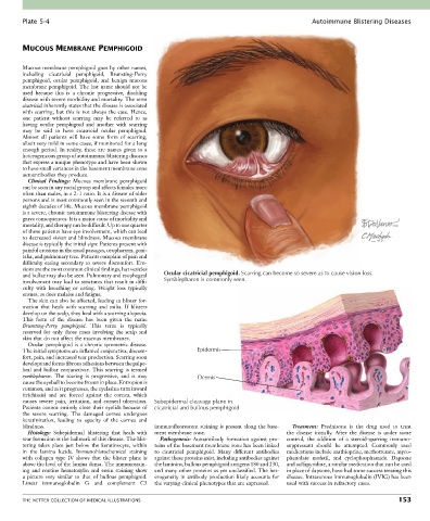
Plate 5-4                                                                                   Autoimmune Blistering Diseases


        MUCOUS MEMBRANE PEMPHIGOID


        Mucous membrane pemphigoid goes by other names,
        including  cicatricial  pemphigoid,  Brunsting-Perry
        pemphigoid,  ocular  pemphigoid,  and  benign  mucous
        membrane pemphigoid. The last name should not be
        used  because  this  is  a  chronic  progressive,  disabling
        disease with severe morbidity and mortality. The term
        cicatricial inherently states that the disease is associated
        with scarring, but this is not always the case. Hence,
        one  patient  without  scarring  may  be  referred  to  as
        having  ocular  pemphigoid  and  another  with  scarring
        may  be  said  to  have  cicatricial  ocular  pemphigoid.
        Almost  all  patients  will  have  some  form  of  scarring,
        albeit very mild in some cases, if monitored for a long
        enough period. In reality, these are names given to a
        heterogeneous group of autoimmune blistering diseases
        that express a unique phenotype and have been shown
        to have small variances in the basement membrane zone
        autoantibodies they produce.
          Clinical  Findings:  Mucous  membrane  pemphigoid
        can be seen in any racial group and affects females more
        often than males, in a 2 : 1 ratio. It is a disease of older
        persons and is most commonly seen in the seventh and
        eighth decades of life. Mucous membrane pemphigoid
        is a severe, chronic autoimmune blistering disease with
        grave consequences. It is a major cause of morbidity and
        mortality, and therapy can be difficult. Up to one quarter
        of these patients have eye involvement, which can lead
        to decreased vision and blindness. Mucous membrane
        disease is typically the initial sign: Patients present with
        painful erosions in the nasal passages, oropharynx, geni-
        talia, and pulmonary tree. Patients complain of pain and
        difficulty eating secondary to severe discomfort. Ero-
        sions are the most common clinical findings, but vesicles
        and bullae may also be seen. Pulmonary and esophageal   Ocular cicatricial pemphigoid. Scarring can become so severe as to cause vision loss.
        involvement may lead to strictures that result in diffi-  Symblepharon is commonly seen.
        culty  with  breathing  or  eating.  Weight  loss  typically
        ensues, as does malaise and fatigue.
          The skin can also be affected, leading to blister for-
        mation  that  heals  with  scarring  and  milia.  If  blisters
        develop on the scalp, they heal with a scarring alopecia.
        This  form  of  the  disease  has  been  given  the  name
        Brunsting-Perry  pemphigoid.  This  term  is  typically
        reserved  for  only  those  cases  involving  the  scalp  and
        skin that do not affect the mucous membranes.
          Ocular pemphigoid is a chronic symmetric disease.
        The initial symptoms are inflamed conjunctiva, discom-  Epidermis
        fort, pain, and increased tear production. Scarring soon
        develops and forms fibrous adhesions between the palpe-
        bral and bulbar conjunctivae. This scarring is termed
        symblepharon.  The  scaring  is  progressive,  and  it  may   Dermis
        cause the eyeball to become frozen in place. Entropion is
        common, and as it progresses, the eyelashes turn inward
        (trichiasis)  and  are  forced  against  the  cornea,  which
        causes  severe  pain,  irritation,  and  corneal  ulceration.   Subepidermal cleavage plane in
        Patients cannot entirely close their eyelids because of     cicatricial and bullous pemphigoid
        the  severe  scarring.  The  damaged  cornea  undergoes
        keratinization,  leading  to  opacity  of  the  cornea  and
        blindness.                                immunofluorescent staining is present along the base-  Treatment:  Prednisone  is  the  drug  used  to  treat
          Histology:  Subepidermal  blistering  that  heals  with   ment membrane zone.     the  disease  initially.  After  the  disease  is  under  some
        scar formation is the hallmark of this disease. The blis-  Pathogenesis:  Autoantibody  formation  against  pro-  control,  the  addition  of  a  steroid-sparring  immuno-
        tering  takes  place  just  below  the  keratinocyte,  within     teins of the basement membrane zone has been linked   suppressant  should  be  attempted.  Commonly  used
        in  the  lamina  lucida.  Immunohistochemical  staining   to  cicatricial  pemphigoid.  Many  different  antibodies   medications include azathioprine, methotrexate, myco-
        with  collagen  type  IV  shows  that  the  blister  plane  is   against these proteins exist, including antibodies against   phenolate  mofetil,  and  cyclophosphamide.  Dapsone
        above the level of the lamina densa. The immunostain-  the laminins, bullous pemphigoid antigens 180 and 230,   and sulfapyridine, a similar medication that can be used
        ing  and  routine  hematoxylin  and  eosin  staining  show     and many other proteins as yet unclassified. The het-  in place of dapsone, have had some success treating this
        a  picture  very  similar  to  that  of  bullous  pemphigoid.   erogeneity in antibody production likely accounts for   disease. Intravenous immunoglobulin (IVIG) has been
        Linear  immunoglobulin  G  and  complement  C3   the varying clinical phenotypes that are expressed.  used with success in refractory cases.


        THE NETTER COLLECTION OF MEDICAL ILLUSTRATIONS                                                                          153