
Page 173 - The Netter Collection of Medical Illustrations - Integumentary System_ Volume 4 ( PDFDrive )
P. 173
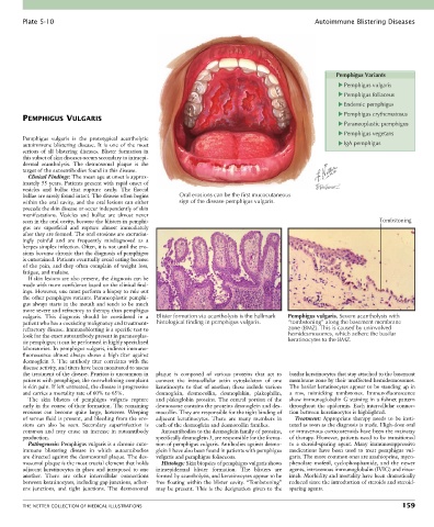
Plate 5-10                                                                                  Autoimmune Blistering Diseases


                                                                                                           Pemphigus Variants
                                                                                                             Pemphigus vulgaris
                                                                                                             Pemphigus foliaceus
                                                                                                             Endemic pemphigus
                                                                                                             Pemphigus erythematosus
        PEMPHIGUS VULGARIS
                                                                                                             Paraneoplastic pemphigus
                                                                                                             Pemphigus vegetans
        Pemphigus  vulgaris  is  the  prototypical  acantholytic
        autoimmune  blistering  disease.  It  is  one  of  the  most                                         IgA pemphigus
        serious  of  all  blistering  diseases.  Blister  formation  in
        this subset of skin diseases occurs secondary to intraepi-
        dermal  acantholysis.  The  desmosomal  plaque  is  the
        target of the autoantibodies found in this disease.
          Clinical Findings: The mean age at onset is approx-
        imately 55 years. Patients present with rapid onset of
        vesicles  and  bullae  that  rupture  easily.  The  flaccid
        bullae are rarely found intact. The disease often begins   Oral erosions can be the first mucocutaneous
        within the oral cavity, and the oral lesions can either   sign of the disease pemphigus vulgaris.
        precede the skin disease or occur independently of skin
        manifestations.  Vesicles  and  bullae  are  almost  never
        seen in the oral cavity, because the blisters in pemphi-                                                         Tombstoning
        gus  are  superficial  and  rupture  almost  immediately
        after they are formed. The oral erosions are excruciat-
        ingly  painful  and  are  frequently  misdiagnosed  as  a
        herpes simplex infection. Often, it is not until the ero-
        sions become chronic that the diagnosis of pemphigus
        is entertained. Patients eventually avoid eating because
        of  the  pain,  and  they  often  complain  of  weight  loss,
        fatigue, and malaise.
          If skin lesions are also present, the diagnosis can be
        made with more confidence based on the clinical find-
        ings. However, one must perform a biopsy to rule out
        the other pemphigus variants. Paraneoplastic pemphi-
        gus always starts in the mouth and tends to be much
        more severe and refractory to therapy than pemphigus
        vulgaris.  This  diagnosis  should  be  considered  in  a   Blister formation via acantholysis is the hallmark  Pemphigus vulgaris. Severe acantholysis with
        patient who has a coexisting malignancy and treatment-  histological finding in pemphigus vulgaris.     “tombstoning” along the basement membrane
        refractory disease. Immunoblotting is a specific test to                            zone (BMZ). This is caused by uninvolved
        look for the exact autoantibody present in paraneoplas-                             hemidesmosomes, which adhere the basilar
        tic pemphigus; it can be performed in highly specialized                            keratinocytes to the BMZ.
        laboratories. In pemphigus vulgaris, indirect immuno-
        fluorescence  almost  always  shows  a  high  titer  against
        desmoglein  3.  The  antibody  titer  correlates  with  the
        disease activity, and titers have been monitored to assess
        the treatment of the disease. Pruritus is uncommon in   plaque  is  composed  of  various  proteins  that  act  to   basilar keratinocytes that stay attached to the basement
        patients with pemphigus; the overwhelming complaint   connect  the  intracellular  actin  cytoskeleton  of  one     membrane zone by their unaffected hemidesmosomes.
        is skin pain. If left untreated, the disease is progressive   keratinocyte to that of another; these include various   The basilar keratinocytes appear to be standing up in
        and carries a mortality rate of 60% to 65%.  desmoglein,  desmocollin,  desmoplakin,  plakophilin,   a  row,  mimicking  tombstones.  Immunofluorescence
          The  skin  blisters  of  pemphigus  vulgaris  rupture     and  plakoglobin  proteins.  The  central  portion  of  the   show immunoglobulin G staining in a fishnet pattern
        early in the course of their formation. The remaining   desmosome contains the proteins desmoglein and des-  throughout  the  epidermis.  Each  intercellular  connec-
        erosions  can  become  quite  large,  however.  Weeping     mocollin. They are responsible for the tight binding of   tion between keratinocytes is highlighted.
        of serous fluid is present, and bleeding from the ero-  adjacent  keratinocytes.  There  are  many  members  in   Treatment:  Appropriate  therapy  needs  to  be  insti-
        sions  can  also  be  seen.  Secondary  superinfection  is   each of the desmoglein and desmocollin families.  tuted as soon as the diagnosis is made. High-dose oral
        common  and  may  cause  an  increase  in  autoantibody   Autoantibodies to the desmoglein family of proteins,   or intravenous corticosteroids have been the mainstay
        production.                               specifically desmoglein 3, are responsible for the forma-  of therapy. However, patients need to be transitioned
          Pathogenesis: Pemphigus vulgaris is a chronic auto-  tion of pemphigus vulgaris. Antibodies against desmo-  to  a  steroid-sparing  agent.  Many  immunosuppressive
        immune  blistering  disease  in  which  autoantibodies     glein 1 have also been found in patients with pemphigus   medications  have  been  used  to  treat  pemphigus  vul-
        are directed against the desmosomal plaque. The des-  vulgaris and pemphigus foliaceous.  garis. The more common ones are azathioprine, myco-
        mosomal plaque is the most crucial element that holds   Histology: Skin biopsies of pemphigus vulgaris shows   phenolate  mofetil,  cyclophosphamide,  and  the  newer
        adjacent keratinocytes in place and juxtaposed to one   intraepidermal  blister  formation.  The  blisters  are   agents, intravenous immunoglobulin (IVIG) and ritux-
        another.  There  are  other  intercellular  connections   formed by acantholysis, and keratinocytes appear to be   imab. Morbidity and mortality have been dramatically
        between keratinocytes, including gap junctions, adher-  free  floating  within  the  blister  cavity.  “Tombstoning”   reduced since the introduction of steroids and steroid-
        ens  junctions,  and  tight  junctions.  The  desmosomal   may  be  present.  This  is  the  designation  given  to  the   sparing agents.


        THE NETTER COLLECTION OF MEDICAL ILLUSTRATIONS                                                                          159