
Page 171 - The Netter Collection of Medical Illustrations - Integumentary System_ Volume 4 ( PDFDrive )
P. 171
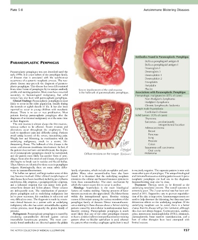
Plate 5-8                                                                                   Autoimmune Blistering Diseases


                                                                                              Antibodies Found in Paraneoplastic Pemphigus
                                                                                                     Bullous pemphigoid antigen II
        PARANEOPLASTIC PEMPHIGUS
                                                                                                     Bullous pemphigoid antigen I
                                                                                                     Desmoglein 1
        Paraneoplastic pemphigus was not described until the                                         Desmoglein 3
        early 1990s. It is a rare subset of the pemphigus family                                     Desmoplakin 1
        of  diseases  that  is  associated  with  the  synchronous                                   Desmoplakin 2
        occurrence of a systemic neoplastic process. The neo-
        plastic disease may precede the diagnosis of paraneo-                                        Envoplakin
        plastic pemphigus. This disease has been differentiated                                      Periplakin
        from other forms of pemphigus by its unique antibody   Severe involvement of the oral mucosa   Plectin
        profile and staining patterns. Most cases have occurred   is the hallmark of paraneoplastic pemphigus.  Associations with Paraneoplastic Pemphigus
        secondary  to  hematological  malignancy,  but  solid                                  Hematologic malignancies (85% of cases)
        tumors have also been with paraneoplastic pemphigus.                                         Non-Hodgkin’s lymphoma
          Clinical Findings: Paraneoplastic pemphigus is most
        likely to occur in the older population, usually during                                      Hodgkin’s lymphoma
        the seventh or eighth decade of life. It has also been                                       Chronic lymphocytic leukemia
        reported  to  occur  in  young  children  with  neoplastic                             Lymph node hyperplasia
        disease.  There  is  no  sex  or  race  predilection.  Most                                  Castleman’s disease
        patients  develop  paraneoplastic  pemphigus  after  the                               Solid tumors  (15% of cases)
        diagnosis of an internal malignancy or at the same time                                      Thymoma
        as their diagnosis.                                                                          Sarcomas—predominantly
          The oral mucosa is almost always the first mucocu-                                                           retroperitoneal location
        taneous  surface  to  be  affected.  Severe  erosions  and                                   Adenocarcinoma
        ulcerations  occur  throughout  the  oropharynx.  This                                         Breast
        leads to significant pain and difficulty eating. Patients
        avoid  eating  because  of  the  severe,  unremitting  pain.                                   Pancreas
        Weight  loss  and  blistering,  in  combination  with  the                                     Lung
        underlying  malignancy,  result  in  a  severe,  life-                                         Prostate
        threatening illness. The hallmark of this disease is the                                       Colon
        severe oral mucous membrane involvement. In fact, if                                         Squamous cell carcinoma
        the patient does not have oral involvement, the diagno-                                        Oral cavity
        sis of paraneoplastic pemphigus should be reevaluated,   Diffuse erosions on the tongue      Melanoma
        and the patient most likely has another form of pem-
        phigus. Soon after the onset of oral disease, the patient’s
        skin begins to break out in vesicles and flaccid bullae.
        These blisters are identical to those seen in pemphigus
        vulgaris.  Histologically,  there  are  some  subtle  differ-
        ences in immunofluorescence.              family of proteins, which include envoplakin and peri-  is routinely negative. The opposite pattern is seen with
          The bullae can spread, and large surface areas of skin   plakin.  Many  other  autoantibodies  have  also  been   most other types of pemphigus. The unique histological
        may become involved. Other clinical morphologies of   found.  It  is  theorized  that  the  underlying  neoplasm   and immunofluorescence staining patterns seen in para-
        skin disease have been described, including an erythema   stimulates the cellular and humoral immune systems to   neoplastic  pemphigus  can  lead  one  to  the  diagnosis.
        multiforme–like eruption, a pemphigoid-like eruption,   form  these  autoantibodies.  The  exact  mechanism  by   Immunoblotting may also be done.
        and  a  lichenoid  eruption  that  can  mimic  both  graft-  which the tumor causes this to occur is unclear.  Treatment:  Therapy  needs  to  be  directed  at  the
        versus-host  disease  and  lichen  planus.  These  variants   Histology:  Acantholysis  is  the  main  histological   underlying neoplastic process. The overall outcome is
        are  infrequently  seen.  The  combination  of  paraneo-  feature on routine staining. Varying amounts of kera-  extremely poor. The 2-year survival rate has been esti-
        plastic  pemphigus  and  an  underlying  malignancy  has   tinocyte necrosis are also appreciated. The blister forms   mated at 10%. Supportive care to prevent superinfec-
        led to poor outcomes; this condition is refractory and   within  the  intraepidermal  space.  Routine  staining   tion of the skin is imperative. Immunosuppressants are
        very difficult to treat. The diagnosis is made by consis-  cannot differentiate among the various members of the   used to help decrease the blistering, but they may have
        tent  clinical  features  in  a  patient  with  an  underlying   pemphigus  family  of  diseases.  Direct  immunofluores-  deleterious effects on the underlying neoplasm. If the
        malignancy who also has serum autoantibodies against   cence staining in these diseases shows a fishnet staining   underlying  neoplasm  can  be  cured,  there  is  a  better
        certain proteins, most frequently the plakin family of   pattern caused by intercellular hemidesmosomal kera-  chance that this disease will go into remission, although
        proteins.                                 tinocyte  staining.  Paraneoplastic  pemphigus  is  much   this does not always happen. Corticosteroids, azathio-
          Pathogenesis: Paraneoplastic pemphigus is caused by   more likely than any of the other pemphigus diseases   prine, intravenous immunoglobulin (IVIG), rituximab,
        circulating  autoantibodies  directed  against  various   to have a positive indirect immunofluorescence staining   plasmapheresis,  bone  marrow  transplantation,  and  a
        intercellular  keratinocyte  proteins.  The  most  com-  pattern when rat bladder epithelium is used, whereas   host  of  other  therapies  have  been  attempted  with
        monly found antibodies are directed against the plakin   the pattern when monkey esophagus epithelium is used   limited success.


        THE NETTER COLLECTION OF MEDICAL ILLUSTRATIONS                                                                          157